Download
1 / 48
530 likes | 830 Views
Emotion & Language. Emotion Classifications. Primary emotions Innate, biologically based. Arise from sensory experience. Same for all humans regardless of culture, and many animals as well. Joy, sadness, fear, disgust, surprise, anger. Secondary emotions

E N D
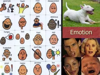
Emotion Classifications • Primary emotions • Innate, biologically based. • Arise from sensory experience. • Same for all humans regardless of culture, and many animals as well. • Joy, sadness, fear, disgust, surprise, anger. • Secondary emotions • Learned & social, dependent on environment. • Pride, shame, embarrassment, anxiety.
Universal (Primary) Emotions • Darwin (1870s) proposed that humans have developed a finite set of basic emotional states, common to all humans. • Ekman and Friesen (1971) studied human cultures around the world and found that facial responses to anger, fear, disgust, happiness, sadness and surprise are universal. • Many animals show similar emotions. • Sensory inputs go directly to the limbic areas.
Isolated New Guinea Tribe “Your friend has come and you are happy.” “Your child has died.” “You are angry and about to fight.” “You see a dead pig that has been lying there for a long time.” Ekman and Friesen (1971)
Secondary Emotions • Secondary emotions (pride, shame, embarrassment, anxiety, etc.) all require higher cortical processing, and are highly dependent on learning and social environment. • Unlike the primary emotions, sensory inputs must first go to the cortex (frontal lobe), and then to the limbic areas.
Secondary Emotions • The frontal cortex is largely responsible. • Cases of Phineas Gage and Elliot – both lost control of appropriate emotion from frontal cortex damage. • The frontal lobe has also been implicated in antisocial behavior disorders, and in schizophrenia, which often features these symptoms.
Cannon-Bard (1927): emotional experience can occur independently of physiological changes. • James-Lange (1884): we perceive emotion in response to physiological changes.
The Papez Circuit • James Papez (1930s) proposed an emotional system in the limbic cortex (identified by Broca in 1878) that ties the neocortex to the hypothalamus, though he had little evidence. • Rabies affects the hippocampus, and causes hyperemotional responses. • Clinical evidence linked anterior thalamus damage to to spontaneous laughing & crying.
The Papez Circuit • Hypothalamus • Governs the expression of emotion thru ANS. • Cingulate cortex • Responsible for emotional experience. • Neocortex • Colors emotions • The bidirectional interconnection of emotional experience and expression areas means the Papez circuit is compatible with both the James-Lange and Cannon-Bard theories.
The Primary Emotions • Damasio (1994) suggested that Ekman’s universal emotions are innate and controlled by the amygdala and cingulate cortex. • Because senses pass thru the thalamus and proceed to the cortex, and back to the limbic areas, it was thought that emotional perception preceded response. • LeDoux suggested a thalamus-limbic shortcut that better matches behavior.
Fear • Fear is good emotion to study since it is innate and easily conditioned to tones with electric shocks, loud noises, etc. • Lesion studies show decerebrate animals still show conditioned fear responses, so the cortex cannot be entirely necessary. • LeDoux showed the amygdala is a crucial part of the fear circuit. The amygdala is also heavily involved in sex, anger and aggression. The hypothalamus is also implicated.
Learned Fear • Kapp (1984) trained rabbits to distinguish two tones, one benign and one with a shock. • Prior to training, central nucleus of the amygdala did not respond. • After training, it responded only to the stimulus associated with the the shock. • LeDoux (1994) showed that visceral responses to the tone disappeared after amygdala lesions.
Learned Fear • fMRI studies of humans show similar responses. • Memories associated with fearful conditions are quickly formed and long lasting. Remember the amygdala is adjacent to the hippocampus. • Amygdala is activated during stimuli associated with punishments, along with cingulate cortex and insular cortex. • AP5 (NMDA antagonist) injections into amygdala inhibit learning of fear responses.
Amygdala • Surgical studies of the amygdala • Bilateral amygdalectomy produces a profound reduction in fear. Stimulation of the lateral amygdala causes fear and violent aggression in cats. • S.M. had bilateral destruction of amygdala from Urbach-Wiethe disease: she lost all recognition of fear expressions, and most expressions of anger. • 1966 Charles Whitman kills 30+ in TX. A walnut-sized glioblastoma multiforme was found compressing his amygdala.
Anger and Aggression • 2 types of aggression • Predatory aggression • Unemotional, for killing, low SNS activation • Usually quiet • Stimulation of the lateral hypothalamus. • Affective/Offensive aggression • Emotional, for show, high SNS activation • Lots of vocalizations • Stimulation of the medial hypothalamus.
Anger and Aggression Summary • The cortex normally inhibits, but can excite. • The amygdala and hypothalamus are both critical. • Androgens/steroids can enhance aggression responses. • Hypothalamus controls SNS. • There is evidence for decreased 5-HT1A and 5HT1B receptor stimulation.
Emotional Asymmetry • The right hemisphere is the major site of visual emotion recognition and is thought to be the major integrator of emotion. • The right inferior frontal area is involved in assessment of audio and visual emotional content. • Some theories suggest the right hemisphere is more responsible for negative emotions, and the left is more responsible for positive emotions.
Emotional Asymmetry Posed smile of pride. RR’ L’L
Emotional Asymmetry • Hallervorden (1902) cut copies of facial pictures in half and joined the same-side halves. • Most people think the pictures of the left face are more emotionally expressive during spontaneous expressions, which would mean the right hemisphere is more responsible for emotions. • The right half of the face is usually more expressive for posed expressions because language is on the left.
Emotional Asymmetry • Type of emotional display matters. • Posed smiles are controlled by the contralateral motor cortices. The right side of the mouth moves first, and generally more than the left. Left stroke patients cannot smile (or smile mostly on the left) for a camera. • Spontaneous smiles of joy seem to originate in the limbic structures, and the smile is more balanced. Left stroke patients have less problem smiling spontaneously.
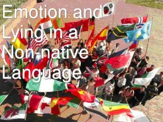
Language • Includes oral, written (books, drawings, etc.), and visual (ASL, flags) language. • Arguably one of the most important classes of human social behavior. No non-verbal human cultures have ever been found. • The most efficient form of communication between members of the species. • Enables learning to be cumulative since we can pass on our knowledge.
Language Terminology • a/an- = without • dys- = malfunctioning • -phasic = speech • -lexic = reading • -graphic = writing • Aphasic = without speech • Dyslexic = poor reading
Language • Studies of the bases of language • Historically limited to observing patients with aphasias or injuries for obvious reasons. • Consequences of strokes, trauma, operations, etc. • Pharmacological tests • Wada test • Electrical cortical stimulation tests • Modern functional imaging techniques have opened a new chapter on language studies and brain studies in general. • MRI, PET, SPECT
History of Language Theories • 1836 Dr. Dax reported that none of his speech-impaired patients had right hemisphere damage (no one paid any attention!) • 1861 Dr. Broca described his patient, Tan, and later 7 more aphasic patients, localizing the area of speech production to the left inferior prefrontal lobe. • Liepmann discovered apraxias are limited to patients with left hemisphere damage. • This lead to a theory of lateralization.
History of Language Theories • 1861 Dr. Pierre Paul Broca described his aphasic patient M. Leborgne, a.k.a. Tan, who was known by the only word he could speak. Dr. Broca discovered during Tan’s autopsy that the left inferior prefrontal cortex, just anterior to the premotor cortex, was damaged. Now named for him, Broca suspected this is involved in language production.
History of Language Theories • 1874 Karl Wernicke studied 10 clinical cases and concluded there is a second language area in the posterior temporal lobe, just behind the primary auditory cortex. He suggested it is involved in language comprehension. This is now known as Wernicke’s Area.
Language Broca’s Area Arcuate fasciculus Wernicke’s Area
Language • From his model, Wernicke predicted: • Broca’s aphasia • Lesions in frontal cortex rostral to base of motor cortex. • Deficits in expressive language, poor repetition and naming. • [Nonfluent (no syntax/grammar) speech, mostly “content” words, but good comprehension,] • Wernicke’s aphasia • Lesions in posterior superior temporal lobe. • Poor comprehension, repetition and naming. • [Deficits in receptive language, nonsensical fluent speech, no semantics.] • Conduction aphasia • Lesions of the arcuate fasciculus. • Fluent speech, good comprehension and naming. • Poor repetition.
Language • 1891 Jules Dejerine • On the basis of 1 patient, concluded that the angular gyrus, just posterior to Wernicke’s area, is responsible for comprehending language-related visual input. • The patient had alexia and agraphia, but no deficits in speech.
Language • Many neurologists of Broca’s and Wernicke’s time refused to believe their localization theories. • 1965 Geschwind re-interpreted and extended their theories to form the Wernicke-Geschwind model of language, including 7 major cortical areas.
Language • We now know the Wernicke-Geschwind model is not perfect: • Lesions to just Broca’s or Wernicke’s areas seldom produce lasting speech deficits. • Severing the arcuate fasciculus produces no permanent aphasia. • Electrical stimulation studies show diffuse and non-specific areas. • Wernicke’s area lesions affect speaking as well.
Language • Studies of dyslexics and Steve Petersen’s PET studies in 1989 lead to the Dual-Route theory of language processing: • Lexical Route • Well-practiced or known verbal responses. • Whole-word recognition. • Nonlexical Route • Novel or complex verbal responses. • Must apply a standard set of rules.
Lexical Processing • Count how many “F”s are in the sentence on the next slide as quickly as you can.
Lexical Processing • FINISHED FILES ARE THE RESULT OF YEARS OF SCIENTIFIC STUDY COMBINED WITH THE EXPERIENCE OF YEARS.
Lexical Processing • The phaomnenel pweor of the hmuan mnid. Aoccdrnig to a rscheearch at Cmabrigde Uinervtisy, it deosn't mttaer in waht oredr the ltteers in a wrod are, the olny iprmoetnt tihng is taht the frist and lsat ltteer be at the rghit pclae. The rset can be a total mses and you can sitll raed it wouthit porbelm. Tihs is bcuseae the huamn mnid deos not raed ervey lteter by istlef, but the wrod as a wlohe.
Dual-Route Theory Received Words Lexical Route (Temporal Lobe) Nonlexical Route (Frontal Lobe) Lexicon Rules (phonetics) Speech Production
Language • Dual-route processing deficits: • Surface dyslexia • Lexical route deficit. • Left lateral temporal lobe lesions. • Inability to pronounce words based on specific memories. • Deep dyslexia or phonological dyslexia • Nonlexical route deficit. • Left frontal lobe damage. • Inability to sound out new words.
Emotional Language • The non-dominant cortex is involved in prosody, the flow and rhythm of speech which often carries emotional content, both in understanding and production. • Aphasias of prosody can render a patient unable to detect or produce the proper emotional content in speech. • Deficits in receptive emotion are often found in the area contralateral to Broca’s area.