Download
1 / 33
330 likes | 535 Views
Interventions for Critically Ill Clients with Acute Coronary Syndrome. Coronary Artery Disease. Includes stable angina pectoris and acute coronary syndromes Ischemia: oxygen supply insufficient to meet requirements of the myocardium

E N D
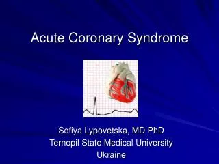
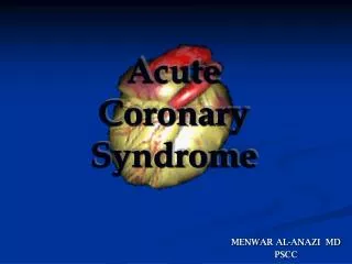
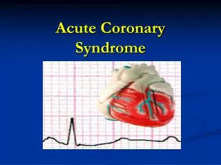
Interventions for Critically Ill Clients with Acute Coronary Syndrome
Coronary Artery Disease • Includes stable angina pectoris and acute coronary syndromes • Ischemia: oxygen supply insufficient to meet requirements of the myocardium • Infarction: necrosis or cell death that occurs when severe ischemia is prolonged and irreversible damage to tissue results
Stable Angina Pectoris • A feeling of “strangling of the chest” • Temporary imbalance between the coronary artery’s ability to supply oxygen and the cardiac muscle’s demand for oxygen • Ischemia limited in duration and does not cause permanent damage to myocardial tissue • Stable and unstable angina
Acute Coronary Syndrome • Atherosclerotic plaque in the coronary artery ruptures, resulting in platelet aggregation, thrombus formation, and vasoconstriction. • Between 10% and 30% of clients with unstable angina progress to having MI within 1 year. • 29% die from MI within 5 years.
Myocardial Infarction • Most serious acute coronary syndrome • Occurs when myocardial tissue is abruptly and severely deprived of oxygen • Dynamic process that does not occur instantly but evolves over several hours
Nonmodifiable Risk Factors • Age • Gender • Family history • Ethnic background
Modifiable Risk Factors • Elevated serum cholesterol • Cigarette smoking • Hypertension • Impaired glucose tolerance • Obesity • Physical inactivity • Stress
Pain Assessment • Discomfort in the chest, epigastric area, jaw, back, or arm is noted. (Rate discomfort on scale of 0 to 10.) • Discomfort is often described as tightness, burning, pressure, or indigestion. • Anginal pain improves with rest and nitroglycerine; MI does not. (Continued)
Pain Assessment(Continued) • Other manifestations include nausea and vomiting, diaphoresis, dizziness, weakness, palpitations, and shortness of breath.
Diagnostic Assessment • Electrocardiogram • Stress test • Myocardial perfusion imaging • Magnetic response imaging • Cardiac catheterization
Acute Pain • Interventions include: • Provide pain relief modalities. • Decrease myocardial oxygen demand. • Increase myocardial oxygen supply.
Pain Management • Nitroglycerine • Morphine sulfate • Oxygen • Position of comfort; semi-Fowler’s position • Quiet and calm environment • Deep breaths to increase oxygenation
Ineffective Tissue Perfusion (Cardiopulmonary) • Interventions include: • Restoration of perfusion to the injured area often limits the amount of extension and improves left ventricular function. • Complete sustained reperfusion of coronary arteries in the first few hours after an MI has decreased mortality.
Thrombolytic Therapy • Fibrinolytics dissolve thrombi in the coronary arteries and restore myocardial blood flow. • Tissue plasminogen activator, APSAC, reteplase • Glycoprotein IIa/IIIb inhibitors
Identification of Coronary Artery Reperfusion • Abrupt cessation of pain or discomfort • Sudden onset of ventricular dysrhythmias • A peak at 12 hours of markers of myocardial damage
Oral Drug Therapy • Aspirin • Beta-adrenergic blocking agents • ACE inhibitors • Calcium channel blockers
Ineffective Coping Interventions • Assess the client’s level of anxiety but allow expression of any anxiety and attempt to define its origin. • Give simple explanations of therapies, expectations, and surroundings, and explanations of progress to help relieve anxiety. • Provide coping enhancement.
Potential for Dysrhythmias • Dysrhythmias are the leading cause of death in most clients with MI who die before they can be hospitalized. • Interventions include: • Identify the dysrhythmias. • Assess hemodynamic status. • Evaluate for discomfort.
Potential for Heart Failure Interventions • Assessment • Monitoring for signs of poor organ perfusion • Hemodynamic monitoring
Cardiogenic Shock • Necrosis of more than 40% of the left ventricle • Tachycardia • Hypotension • Blood pressure < 90 mm Hg or 30 mm Hg < client’s baseline • Urine output < 30 mL/hr (Continued)
Cardiogenic Shock (Continued) • Cold, clammy skin • Poor peripheral pulses • Agitation, restlessness, confusion • Pulmonary congestion • Tachypnea • Continuing chest discomfort
Medical Management • Pain relief and decreased myocardial oxygen requirements through preload and afterload reduction • Intravenous morphine • Oxygen, intubation, ventilation • Intra-aortic balloon pump • Immediate reperfusion
Potential for Recurrent Symptoms and Extension of Injury Interventions • Percutaneous transluminal coronary angioplasty (PTCA) • Coronary artery bypass graft surgery (CABG)
Percutaneous Transluminal Coronary Angioplasty • Monitoring for acute closure of the vessel, bleeding from the insertion site, reaction to dye, hypotension, hypokalemia, and dysrhythmias • Long-term nitrate, calcium channel blocker, and aspirin therapy • Beta blocker and ACE inhibitor if MI • Infusions of GPIIa/IIIb inhibitors
Coronary Artery Bypass Graft Surgery • Postoperative care in cardiopulmonary bypass • Management of fluid and electrolyte imbalance, hypotension, hypothermia, hypertension, bleeding, cardiac tamponade, altered levels of consciousness, and pain
Transfer from the Special Care Unit • Ventilation provided for 3 to 6 hours postoperatively • Supraventricular dysrhythmias commonly occur • Sternal wound infections • Mediastinitis • Postpericardiotomy syndrome
Other Interventions • Minimally invasive direct coronary artery bypass • Transmyocardial laser revascularization • Off-pump coronary artery bypass • Robotics
Health Teaching • Smoking cessation • Diet control • Complementary and alternative therapies • Physical activity • Sexual activity (Continued)
Health Teaching (Continued) • Blood pressure, blood glucose control • Cardiac medications • Self-monitoring; seeking medical assistance if needed