Download
1 / 42
420 likes | 673 Views
A 25 y/o pregnant woman with acute heart failure. Ri 曾渥然 /Ri 楊智勝 /CR 康庭瑞 /VS 鄭淳心. Brief History. 25 y/o female patient GA 35 + weeks with twin pregnancy BL: 155 cm BW: 47/62 kg Past history: denied. OB/GYN history. G2P0AA1 LMP: 94.4.9 EDC: 95.1.16 Prenatal condition GDM: negative

E N D
A 25 y/o pregnant woman with acute heart failure Ri曾渥然/Ri楊智勝/CR康庭瑞/VS鄭淳心
Brief History • 25 y/o female patient • GA 35+ weeks with twin pregnancy • BL: 155 cm • BW: 47/62 kg • Past history: denied
OB/GYN history • G2P0AA1 • LMP: 94.4.9 • EDC: 95.1.16 • Prenatal condition • GDM: negative • VDRL: non-reactive • HIV: negative • Blood type: B, Rh(+) • SMN carrier (-)
Prenatal examination • 94.11.14 (31+2 wks) • Urine sugar(-), Urine protein(-) • 59kg, 91/83 mmHg • 94.11.30 (33+5 wks) • 62kg, 121/97 mmHg • Urine sugar(-), Urine protein(2+) • 94.12.14 ( 35+5 wks) • 66kg, 130/95 mmHg • Urine sugar(-), Urine protein(4+)
Clinical symptoms and signs • Exersional dyspnea for one week • Orthopnea • Vital signs • 36.5/129/20, BP:135/108mmHg • PE • Neck: JVE(+) • Chest: bilateral basal crackles • Heart: RHB with systolic murmur • Abd: ovoid, normactive BoS • Ext: edema (+)
Cardiac Echo (12.14) • Moderate to severe MR • Moderate PR & TR • LVEF: 43% • LV dilatation (47mm) • Pulmonary hypertension • PG = 50mmHg • Small amount pericardial effusion
Impression • Twin pregnancy at 35th week s/p emergent Cesarean section • Preeclampsia • Maternal heart failure, suspected cardiomyopathy
Anaesthesia Records • Arterial line • Induction agents • Atropine • Etomidate • Succinylcholine • Under ETGA (6:50PM) • Inhalation of isoflurane • Cis-atracurium for m. relaxant • Midazolam for sedation • Fentanyl for pain control • Central venous catheter
Delivery time: 7:03PM • Twin A (vertex extraction) • BBW:2016gm • Apgar score: 5 8 • Fetal anomaly: nil • Twin B (breech extraction) • BBW:2344gm • Apgar score: 5 8 • Fetal anomaly: nil • Placenta: intact • EBL: 500ml
4FI treatment course(II) • Medication • Albumin 2btls IV x 3 days • Lasix: 1 amp TID • Perdipine titration • MgSO4 in D5W • Inderal(10) 1tab BID • Ventilator settings • Mode: SIMV + PS • FiO2:35% • PS/PEEP:8/4 • SpO2:96% • Extubation at 12.16_10:45AM
12/14 Send to 4 FI • 12/16 ETT was removed • 12/17 Transfer to general ward • 12/21 Discharge
Followed up cardiac echo • 12/16 • LVEF:62% • LVEDD:49mm • Moderate MR • TR, PG:44mmHg • 12/20 • LVEF:48% • LVEDD:55mm • Moderate to severe MR • TR, PG:36mmHg
Discussion • Maternal hemodynamic change during pregnancy • Recognition and management of peripartum cardiomyoparthy
Maternal hemodynamic change during pregnancy • Heart • Cardiac output • MAP and SVR • Venous return • Hemodynamic assessment
Heart • Displacement of diaphragm - left and upward - apex is moved laterally • Expand of blood volume - LVEDD increase - LA/RA diameter increase
Cardiac output • Position - Increased: left lateral - Decreased: sitting, supine
MAP and SVR • Decreased SVR - Elevated progesterone level - NO, prostaglandin, ANP • Refractory to hypertensive effects of angiotensin II
Venous pressure • Elevated in lower extremities • Edema • Varicose veins • Deep vein thrombosis
Hemodynamic assessment • By arterial lines and Swan-Ganz catheterization
Effect of labor and immediate puerperium • Elevated MAP and CO - uterine contraction - pain and apprehension - position
Diagnosis and evaluation of cardiac disease in pregnancy • Detailed historical information • Physical examination: • murmur(≧ grade 3/6) or radiate to carotid→ pathologic • JVE, peripheral cyanosis, clubbing, pulmonary crackle • Echocardiography
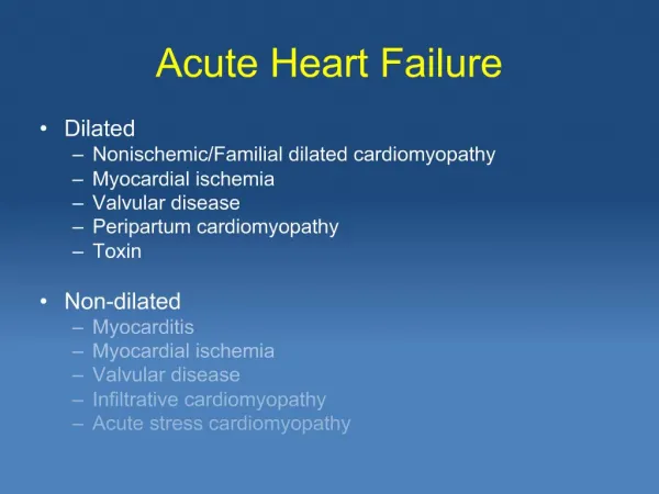
The possible etiology of heart failure in this patient • Previously undiagnosed congenital or rheumatic disease • Embolic disease or ischemic heart disease • Cardiomyopathy • Previously undiagnosed valvular disease
Maternal death due to cardiac disease Br J Anaesth 2004;93, 428-39
Peripartum cardiomyopathy • Incidence: 1:1500~1: 4000 • It is associated with older maternal age, greater parity, black race and multiple gestation… • The etiology remains unclear.
Clinical definition of peripartum cardiomyopathy • Heart failure within last month of pregnancy or five months postpartum • Absence of prior heart disease • No determinable cause • Strict echocardiographic indication of LV dysfunction • Ejection fraction < 45% And/or • Fraction shortening < 30 % • End-diastolic dimension >2.7 cm per m2 BSA
The recognition of peripartum cardiomyopathy • The diagnosis as a challenge • S/S that should raise the suspicion of HF, including paroxysmal nocturnal dyspnea, chest pain, new regurgitant murmur, pulmonary crackles, JVE, hepatomegaly… • EKG: usually NSR or sinus tachycardia • A high index of suspicion and low threshold of cardiac echo
Treatment of peripartum cardiomyopathy • Salt restriction and the use of diuretics • If systolic dysfunction, the use of vasodilators to reduce afterload • ACEI→ teratogenicity, neonatal anuric renal failure (contraindicated) • Hydralazine: the drug of choice prepartum • Atrial arrhythmias should be treated with digoxin. • Class 3(amiodarone) and class 4(verapamil) agents have side effects: fetal hypothyroidism, premature delivery, fetal bradycardia…
Treatment of peripartum cardiomyopathy (Cont.) • Patient with poor cardiac output (LVEF<35%) • Anticoagulation is indicated for the risk of thromboembolism. • Unfractionated or LMW heparin are the choice during pregnancy. • The use of β- blocker • Carvedilol for dilative cardiomyopathy • The application in peripartum cardiomyopathy is unclear.
Cardiomyopathy during pregnancy • Delivery of the fetus ↓ the hemodynamic stress on the heart. • The mode of delivery • Based on obstetric indication • Effective pain management is necessary.
Anesthetic consideration in this patient • In patients undergoing GA, the principles are as for any patient with cardiac failure. • Maintenance of normal to low heart rate to ↓O2 demand • Prevention of large swings in BP • The hypertensive response to intubation can be obtunded by the use of alfentanil.
Anesthetic implication for stage-C heart failure • Two principal cardiovascular events: • Myocardial depression • Peripheral vasodilatation • The patients with impaired LV function are dependent on • the preload • increased sympathetic tone
Anesthetic consideration in this patient (Cont.) • Careful monitoring of fluid balance • Arterial line • CVP line • Early critical care referral for unstable patient • Critical patients will require Swan-Ganz monitoring, artificial ventilation and inotropic support.
Severe myocardial dysfunction • The use of IABP or a left ventricular assisted device needed as a bridge until myocardial recovery or cardiac transplantation is performed.
The prognosis of peripartum cardiomyopathy • The mortality rate: 25~50% • Death due to progressive CHF, arrhythmia, or thromboembolism. • Within 6 months: • Half of patient have resolution of LV dilation. • If not, 85% will die within the next 4~5 years.