Download
1 / 19
300 likes | 617 Views
Manchester Biomedical Research Centre. Age-related macular degeneration and the Y402H polymorphism in complement factor H. Tony Day. Simon Clark. Progression of AMD. Age-related maculopathy Age-related macular degeneration Atrophic Neovascular.

E N D
Manchester Biomedical Research Centre Age-related macular degeneration and the Y402H polymorphism in complement factor H Tony Day Simon Clark
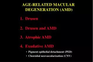
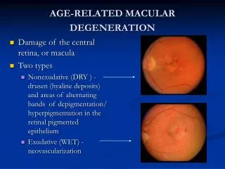
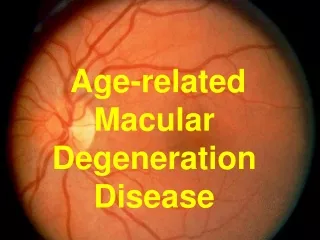
Progression of AMD Age-related maculopathy Age-related macular degeneration Atrophic Neovascular Soft drusen + pigmentary changes in RPE
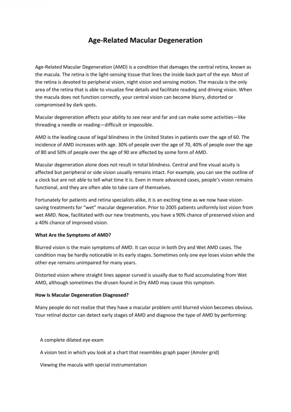
AMD is a condition that primarily affects RPE cells in the macula • Why particularly the macula? • Most metabolically active • Light damage
Ageing changes within the RPE • Intracellular accumulation of lipofuscin • Autofluorescentmaterial that accumulates in lysosomes • Contains lipid and protein • Formed from incompletely degraded photoreceptor outer segments and autophagy • Induces free-radical formation
Accumulation of extracellular material derived from RPE Drusen Diffuse thickening of Bruch’s membrane RPE cells Thickening of Bruch’s Membrane (basal linear deposits) Extracellular material disrupts transport between RPE and choroid and thereby increases RPE stress
Genetic susceptibility Environmental factors Ageing retina and progression to AMD High metabolic activity, light, oxygen resulting in free radical formation and RPE dysfunction Inflammation and accumulation of intracellular and extracellular debris Critical point reached Death of retinal/RPE cells Atrophy Choroidal neovascularisation
What predisposes to AMD apart from ageing? • Genetic risk factors: Major risk factors • Complement factor H (especially Y402H polymorphism) • ARMS2/HTRA1 locus (Chr 10q26) Intermediate risk factors • Complement factor 2/complement factor B locus • Complement factor 3 Minor risk factors • ABCA4 • ApoE Decreased risk CFHR1/CFHR3 deletion • Environmental influences • Smoking ↑ • Abdominal adiposity ↑ • Anti-oxidant intake ↓ • Omega-3 fatty acid intake ↓
Complement cascade * Implicated in AMD * * * Cellular damage, phagocytosis, inflammation
Functions of Complement Factor H (CFH) • There is tick over C3b is deposition on host surfaces which can amplify and lead to activation of alternative pathway unless suppressed • CFH is a soluble protein mainly produced by the liver that binds to polyanionic structures (such as glycosaminoglycans (GAGs)) on host surfaces and prevents C3b mediated complement activation
Structure of CFH CFH consists of 20 complement control protein (CCP) modules Y402H polymorphism is a major genetic risk factor for AMD 402 residue contributes to GAG binding site (Prosser et al., J. Exp Med 2007)
Glycosaminoglycans (GAGs) Heparan sulfate (HS) proteoglycans Heparin a highly sulfated form of HS made specifically by mast cells Types of GAGs
Drusen RPE Bruch’s membrane Immunolocalisation of CFH in human macula tissue (OX23/24 Abs)
Hypothesis and research question • Hypothesis – differential binding of 402H and 402Y forms to GAGs explains the association of the polymorphism with AMD (still debate as to whether this is the functional change associated with this genetic locus) • Question – Do the 402H and 402Y forms show differential binding to (normal) macular tissue
402H CCP6-8 (disease associated) 6 H 8 H 402Y CCP6-8 6 Y 8 Y Methodology Full length CFH 402H Full length CFH 402Y • 402H and 402Y CCP6-8 (recombinant) or CFH (full-length) were fluorescently-labelled (H - red) (Y - green) and applied in pairs to frozen sections of “normal” post mortem human macula tissue (age 46-90) • Relative levels of binding of the 402H and 402Y forms were calculated after measuring fluorescence (and subtracting background fluorescence) • In some experiments tissue sections treated with GAG degrading enzymes prior to application of fluorescently-labelled proteins
merge merge 402H 402H 402Y 402Y RPE 100 µm CCP6-8 Bruch’s membrane flCFH Results402H and 402Y CCP6-8 and full-length CFH CCP6-8 200 * 150 Relative fluorescence (arbitrary units) 100 50 402H 402Y 402H 402Y 0 RPE Bruch’s membrane Note similar pattern in choroidal vessels flCFH 100 ** 80 60 Relative fluorescence (arbitrary units) 40 20 402H 402Y 402H 402Y 0 RPE Bruch’s membrane Control experiments: Binding unaffected by genotype of tissue Results unaffected by exchanging fluorophores Unlabelled proteins effectively competed for binding * P = 0.0005, ** P = 0.009
402H and 402Y (CCP6-8) binding sites are predominantly heparan sulphate (HS) with a smaller amounts of dermatan sulphate (DS) Relative binding of 402H and 402Y from previous slide No evidence of interactions with hyaluronan or chondroitin sulfate
Effects of heparinise and chondroitinase B digestion on binding of full length 402H and 402Y and CFH antibody labelling
Binding of CCP6-8 Y and H variants to selectively desulfated heparin(Heparin is a highly sulfated form of HS)
Conclusion • 402H form of CFH binds poorly to Bruch’s membrane and choroidalstructures compared to 402Y form • CFH binds particularly to HS and the differing affinities of 402H and 402Y forms and dependent upon HS sulfation patterns • Suggests a possible disease mechanism….. • Supports the Y402H amino acid substitution being the genetic alteration that predisposes to AMD. 402Y form localises to Bruch’s membrane and thereby protects against complement activation on the membrane 402H form localises poorly to Bruch’s membrane and this may lead to complement over-activation, inflammation and eventually AMD