Download

1 / 41

E N D
Anatomy of Hypothalamus KamalSalih
Galen of Pergamon reported fundamental discoveries in the anatomy of the third ventricle region, including the location of the pituitary gland inside the sellaturcica embodied in a vascular network, the rete mirabilis, and observed nerves adjoining the "soft flesh" in the neck, i.e. the thyroid gland
William His 1895 • The first to use the term hypothalamus
Auguste-Henri Forel(1848-1931) • Swiss neuroanatomist, psychiatrist, and entomologist. • He gave such a precise description of the hypothalamus that one of its regions - Nucleus hypothalamicus - is called Campus Foreli, or Forel's body, in his honour
Walter Rudolph Hess • In 1928, The first to report "affective responses" to hypothalamic stimulation
Boundaries and Divisions • Connections • Functions of the Hypothalamus • Blood Supply
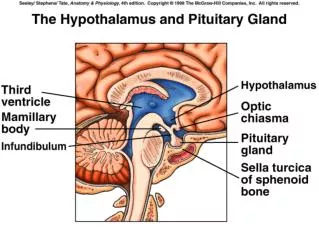
The hypothalamus is the area of the diencephalon ventral to the hypothalamic sulcus
Weight • It weighs about 4 g and comprises 0.3 to 0.5 percent of brain volume.
It is limited anteriorly by the lamina terminalis and is continuous posteriorly with the mesencephalon.
On its ventral surface, caudal to the optic chiasma, the hypothalamus narrows to a small neck, the tuber cinereum.
The median eminence blends into the infundibular stalk, which is continuous with the posterior lobe of the pituitary gland (hypophysis).
In coronal sections, the hypothalamus is bordered medially by the third ventricle and laterally by the subthalamus
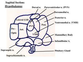
The fornix divides the hypothalamus into:- • Lateral region • Medial region
The lateral region • The lateral region contains mainly longitudinally oriented fibers of the medial forebrain bundle (which connects the septal area, hypothalamus, and midbrain tegmentum), among which are scattered neurons of the lateral hypothalamic nucleus.
The medial region • The medial region has a cluster of nuclei organized into four major groups.
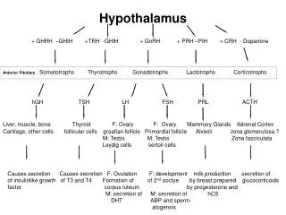
Nuclear groups of medial part • In a rostrocaudal orientation these nuclear groups are as follows: • Preoptic • Suprachiasmatic (supraoptic) • Tuberal • Mamillary
Boundaries and Divisions • Preoptic Region • Suprachiasmatic (Supraoptic) • Region Tuberal Region • Mamillary Region
The Medial Preoptic Nucleus • The medial preoptic nucleus contains neurons that elaborate gonadotropic releasing hormone which reaches the anterior pituitary gland via the tuberoinfundibular tract.
The Medial Preoptic Nucleus • It is related to reproduction, eating, locomotion, and sexual arousal.
The Medial Preoptic Nucleus • Eating • Locomotion • Sexual arousal
The Medial Preoptic Nucleus • The medial preoptic nucleus is referred to as the sexually dimorphic nucleus.
The Medial Preoptic Nucleus • It is twice as large in young males compared to females, probably because gonadotropin release in males is constant, whereas it is cyclic in females.
The Medial Preoptic Nucleus • The difference in size may also explain the reported greater sexual arousal to erotic stimuli experienced by men.
The Medial Preoptic Nucleus • Functional magnetic resonance imaging (fMRI) studies have shown greater activation in the preoptic region in men compared to women when viewing erotic films.
Suprachiasmatic (Supraoptic) Region • Located above the optic chiasma, this nuclear group contains the • Supraoptic nucleus • Paraventricular nucleus • Anterior hypothalamic nucleus • Suprachiasmatic nucleus.
Suprachiasmatic (Supraoptic) Region The supraoptic nucleus is located above the optic tract, whereas the paraventricular nucleus is dorsal to it, lateral to the third ventricle.
Magnocellular Neurons • The supraoptic and paraventricular nuclei contain magnocellularsecretory neurons.
Supraoptic& Paraventricular Axons • Axons of the supraoptic and paraventricular nuclei course in the pituitary stalk to reach the posterior lobe of the pituitary (hypothalamoneurohypophyseal system), transporting neurosecretory material elaborated in these nuclei and stored in axonal swellings within the posterior lobe.
Suprachiasmatic (Supraoptic) Region • The neurosecretory material consists of vasopressin ADH and oxytocin.
Suprachiasmatic (Supraoptic) Region • There is evidence to suggest that the supraoptic nucleus elaborates mainly ADH, whereas the paraventricular nucleus elaborates mainly oxytocin.