Download
1 / 31
430 likes | 2.43k Views
Medical parasitology lab. Staining of Parasites 2. Infect human and most mammals. The infective stage is oocyst containing sporozoites measuring 4-6µ in diameter. The diagnostics stage is oocyst containing 4 sporozoites. Diagnosis: Detecting oocyst in stool. Acid-fast stain.

E N D
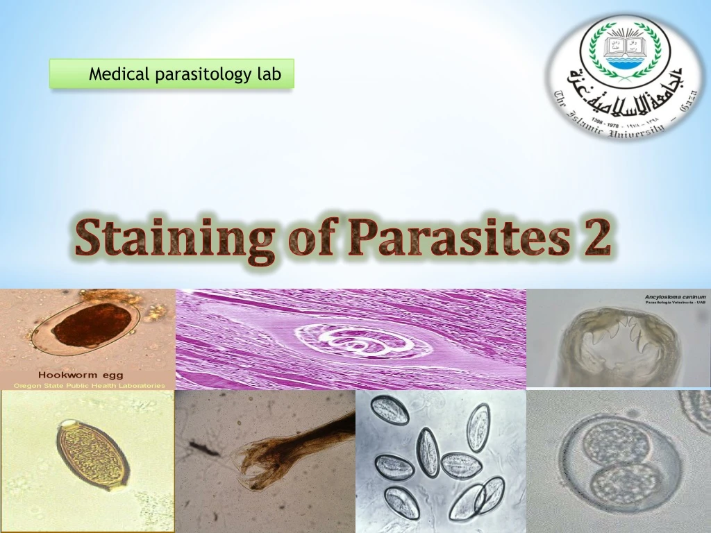
Medical parasitology lab Staining of Parasites 2
Infect human and most mammals. • The infective stage is oocyst containing sporozoites measuring 4-6µ in diameter. • The diagnostics stage is oocyst containing 4 sporozoites. • Diagnosis: • Detecting oocyst in stool. • Acid-fast stain Cryptosporidium parvum
Isosporiasis is a human intestinal disease caused by the parasite Isospora belli. • The coccidian parasite Isospora belli infects the epithelial cells of the small intestine, and is the least common of the three intestinal coccidia that infect humans. • Infection causes acute, non-bloody diarrhea with crampy abdominal pain, which can last for weeks and result in malabsorption and weight loss. In immunodepressed patients, and in infants and children, the diarrhea can be severe. Eosinophilia may be present. • Diagnosis: • Acid- fast stain Isospora belli
Cryptosporidium parvum_ wet mount Notice how easy it would be to miss this on a fecal ova and parasite examination.
Oocysts are elongated ellipsoidal in shape, one sporoblast or two sporocysts appear inside Oocyst wall is thin and colorless, unexperiencedmicroscopistmay miss it easily
Cryptosporidium parvumoocyst Isospora belli oocyst Oocysts in clinical specimens may be difficult to identify with out special staining. Modified acid-fast (partial acid-fast) stains are recommended for identifying these organisms. This test detects coccidian parasites (Cryptosporidium, Cyclospora, or Isospora belli) in stool. It is used to evaluate chronic diarrhea.
The oocysts absorb the red from the carbol-fuchsin stain and may appear in a range of colors from pink to dark purple with bright red being typically seen. • The background material typically stains blue or light red. • Specimen Collection Concentrated sediment of fresh or formalin preserved stool may be used. Other types of clinical specimens such as bile , duodenal fluid, pulmonary fluid (induced sputum, bronchial washings, biopsy specimens may also be used to stain for organisms. Principle of acid fast stain
procedure 1 2 3 1-carbol-fuchsin for 3-5 mint 2-rinse by tap water 3- add acid alcohol as decolorizer for 1mints 4- rinse by tapewater 5- add methylene blue as counter stain for 1 mint Rinse by tapewater then examine under oil immersion. 4 5
The iron hematoxylin stain reveals excellent morphology of the intestinal protozoa. • Iron hematoxylin was the stain used for most of the original morphological descriptions of intestinal protozoa found in humans . • Under oil immersion power (1,000),one can examine the diagnostic features used to identify the protozoan parasite. Iron Hematoxylin Stain
Place approximately 50 ml of deionized water in separate coplin jar.
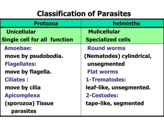
General characters • Non segmented • Sex are separate Male is smaller than female and its posterior end is curved ventrally. Female are either • Larviparousor viviparous: giving birth to larvae • Oviparous: laying egg • Oviviviparous: laying eggs which contain larvae and which hatch out immediatly Nematodes = roundworm
Tissue Nematodes Trichinellaspiralis Intestinal Nematodes Entrobiusvermicularis Trichuristrichiura Ancylostomaduodenale Strongyloidesstercoralis Ascarislumbricoides
Trichinellaspiralis • Adult inhabit the small intestine of the rats, pigs and human. Both males and females lie freely in the lumen of the intestine of pigs, rats and human. • Fertilized female only penetrate the mucosa where the larviposit, they do not lay eggs.
Eggs Adults Entrobiusvermicularis
Young and mature worms are present in small intestine (at terminal ileum till fertilization).
Egg Morphology : • 20-50u, transparent with double walled shell. • Oval, It may show one side convex and the other flat. • Shell: double layered, thick, colorless. • Embryo stage of development varies may be unembryonated, embryonated, mature. • Microscopically examined the slide under low power. Reduced light is recommeneded as the eggs will appeear colorless, making them difficult to detect under high light intensity .
These cells are readily differentiated from parasitic forms because they lack internal structures. Starch artifact can confused with egg of Entrobiusvermicularis
The female is 0.88-1.3 cm long, it has a long tapering tail resembling 1/3 its length, its straight.Gravid females are present at lower rectum where they lay ova at perianal region around anus (oviparous). The tail of female pointed resembles pinhead The male is shorter than female (2-5 mm) the tail is curved strongly to ventral side, and has a single spicule.
Adhesive tape • Use gloves !(pinworm eggs are infectious!) • Prepare the tape then Take sample on peri-anal skin in the morning • Stick tape on microscope slide • Place a drop of Xylene on the edge of the tape to remove airbubbles Adhesive tape method for the detectionof pinworm eggs
Adult inhabit the large intestine in the caecum of man. • The adult male smaller than female, male 3.4-4.5 cm, female 4-5 cm. • Its commonly called whip worm because of the shape of this worm (anterior thin and posterior thick).
Trichuristrichiuraeggs have distinct shape. (oviparous) • Shell:smooth,yellow brown color due to bite contact. • Hyaline plug at each pole. • Diagnosis: • Stool examination to detect eggs.