Download
1 / 58
620 likes | 767 Views
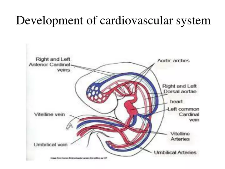
Development of cardiovascular system. Primitiv e blood circulation . Heart development ( dev. Of heart tube, septa and valves ) Aort a l arches a nd their deriv a t ives . Fet a l blood circulation . C ardiovas c ul a r syst e m malformations. Vessels development: (from week 3)

E N D
Primitivebloodcirculation. • Heart development (dev. Of heart tube, septa and valves) • Aortal arches and their derivatives. • Fetal blood circulation. • Cardiovascular system malformations.
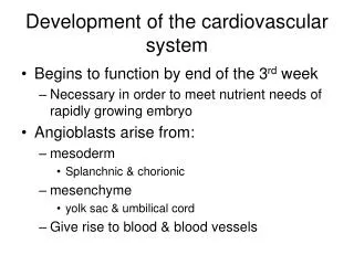
Vessels development: (from week 3) hemangiogenesis - blood islands (insulae sanguinae) DAY 15 – 16 in extraembryonic mesoderm of - yolk sac (vasa omphalomesenterica /vitellina/), - connecting stalk and placenta (vasa umbilicalia) DAY 17 – 18 in mesenchyme ofembryo
Groups of angiogenic cells in mesenchyma ectoderm mesenchyme endoderm Blood islet angioblasts hemoblasts primitive erythrocytes primitive endothelium Differentiation of mesenchymal cells angiogenic cells: - angioblasts endothelium (at the periphery of blood islets) - hemoblasts primitive erythrocytes (in the center of blood islets)
angiogenic cells form a "horseshoe-shaped" space between somatic and splanchnic layer of mesoderm = pericardial cavity. Two endothelial tubes arrise in splanchnic mesoderm. The ventral portion with tubes forms the cardiogenic area twoheart tubes, while the lateral portions form the dorsal aortae.
Vitelline, umbilical and intraembryonic vessels fuse together and form primitive blood circulation (NOfetal blood circulation!)
arterial end of heart tube aortic sac dorsal aortae
Veins entering venous end of herat tube: v. cardinalis communis
branchial arteries 1-6 Desc.aorta Truncus arteriosus Asc.aorta Sinus venosus Saccus aorticus v. precardinalis sin. v. cardinalis com.sin. v. postcardinalis sin. Vessels + cor tubulare Yolk sac aa.omphalomesentericae a. mesenterica sin. aorta dorsalis Connecting stalk v. umbilicalis sin. v. omphalomesenterica sin. a. iliaca com.sin. a. umbilicalis sin. from a. iliaca int.
Heart development Space between somatic (parietal) and splanchnic (visceral) mesoderm = perikardial cavitywith 2 endotelial tubes in splanchnic mesoderm – heart (ventromedially) 2 endotelial tubes – dorsal aortae (ventrolaterally on arterial end of heart tubes).
Heart tubes(day 18-19) • in mesenchyme of splanchnopleura – 2 endothelial tubes • As a result of embryo flexion: • separation of yolk sac • heart tubes fusion COR TUBULARE SIMPLEX (contractions – day 23, sono registration – week 4)
Histogenesis of heart tube wall COR TUBULARE SIMPLEX– temporarily suspended on mesocardium dorsale (4). Visceral mesoderm (splanchnopleura, 5) propagates and forms myoepicardial coat myocardium (b) + epicardium (a). Below endothelium (d) – layer of cardiac jelly subendocardiac connective tissue (c). a b c d
Heart tube(s) Pericardial cavity
Bulboventricular loop Dorso- cranially Ventro- caudally to the right ventral viewlateral view
Heart tube Cor tubulare simplex Cor sigmoideum uniloculare Cor quadricameratum Truncus arteriosus + saccus aorticus Bulbus cordis Ventriculus Atrium Sinus venosus vv. cardinales communes vv. umbilicales vv. vitellinae
Septum atrioventriculare A/ Endocardial cushions – fromdorsal (3) and ventral (2) wall of atrioventrikular canal. Grow against each other and seperate right and left AV canal (4, 5). B/ Lateral interventricular cushions – bicuspid + tricuspid AV valve.
Ventriculus Septum interventriculare Grows from apex cordis craniallly to AV septum • - membranózní část septa • - IV septum
Atrium • Septum atriorum • septum primum s ostium primum (obliteration); ostium secundum • septum secundum with foramen ovale
Septum primum Grows from dorsocranial wall – foramen primum (caudally), closes later, and foramen secundum (above) appears by cell apoptosis S1 – septum primum, O1 – foramen secundum
In septum primum by cell apoptosis foramen secundum will arrise S1 – septum primum, SS – septum spurium, O1 – foramen primum, EC – endocard. cushion, Perf – perforation of foramen secundum, SAO – sinoatrial orifice,
Septum secundum • semicircular fold, does not reache endocardial cushions; • covers foramen secundum in septum primum and by its free lower margin surrounds foramen ovale S1 – septum primum, S2 – septum secundum, SS – septum spurium, O1 – foramen secundum, FO – foramen ovale, EC – endocard. cushion, LVV – left venous valve
before and after birth S1 – septum primum (valvula foraminis ovalis), S2 – septum secundum, SS – septum spurium, O1 – foramen secundum, FO – foramen ovale, EC – endokardový polštářek, Perf – perforace, SAO – sinoatriání orificium, LVV – levá venózní chlopeň Blood from v. cava under pressure flows from the right atrium into the left.
1. Vena cava superior2. Venae pulmonales3. Atrium sin.4. Atrium dx.5. Septum primum6. Septum secundum7. Primitive left atrium8. Primitive right atrium9. Valve of vena cava inferior10. Valve of sinus coronarius11. Sinus venosus
Sinus venosus vv. cardinales communes vv. umbilicales vv. vitellinae Left veins obliterate and - left part of sinus venosus sinus coronarius - right part of sinus venosus part of right atrium wall
Sinus venosus + atria On the right side: v. cava sup. from v. cardin. comm. dx.+ v. precardin. dx. v. cava inf. (posthepatic part) from v. omphalomes. dx. On the left side: veins obliterate and give rise to sinus coronarius Truncus arteriosus atrium • Sinus venosus: • transvesal part • R + L horns: • v. cardinalis comm. • v. umbilicalis • v. omphalomesenterica
Truncus arteriosus + saccus aorticus • cranial part of bulbus cordis separates into: • 2 aortic roots with 6 pairs of aortic arches
Bulbus cordis • cranial – truncus arteriosus • middle – conus arteriosus -caudal – part of ventricle wall
Bulbus cordis – participate in ventricle wall; in RV - conus arteriosus, in LV– sinus aortae.
Bulbus cordis a truncus arteriosus a pair of opposing ridges appear in the walls of the bulbus cordis and truncus arteriosus. These ridges twist around each other, forming a spiral course of the aortico-pulmonary septum. This septum divides the bulbus cordis and truncus arteriosus into two channels, the aorta and the pulmonary artery.It also participates in the closure of the interventricular foramen
Septum aortopulmonale 1. Aorta2. a. pulmonalis sin.3. Truncus pulmonalis4. Septum interventriculare(svalová část)5. pravá komora6. membranózní část septum interventriculare
1. Septum aorticopulmonale2. Pulmonálníchlopeň3. A. pulmonalis4. Aortální chlopeň5. Aorta
1. Internal carotid artery2. External carotid artery3. Common carotid artery4. Right subclavian artery5. Arch of aorta6. Brachiocephalic artery7. Ductus arteriosus8. 7th intersegmental artery9. Pulmonary artery10. Carotid duct11. Obliterated right dorsal aorta
At birth, the circulation of the fetal blood through the placenta is stopped and the lungs begin to function. The foramen ovale, ductus arteriosus, ductus venosus and umbilical vessels subsequently obliterate and transform into corresponding ligaments.
Congenital malformations in CVS(the most frequent) • With left – right shunt (without cyanosis) atrial septum defect ventricular septum defect ductus arteriosus apertus (= patens, = persistens) • With right – left shunt (with cyanosis) Fallot tetralogy transposition of great vessels truncus arteriosus (common aorticopulmonal canal) tricuspid valve atresia • Without shunt coarctation of aorta aortic stenosis pulmonary stenosis dextrocardia (+situs inversus) ectopia cordis
Atrial Septal Defects a group of common congenital anomalies defects occuring in a number of different forms and more often in females. • patent foramen ovale • left-right shunting
Ventricular Septal Defect The Ventricular Septal Defect occurs in the interventricular septum, and is more frequent in males that females. left-right shunting
Patent Ductus Arteriosus occurs commonly in preterm infants, can close spontaneously (by day three in 60% of normal term neonates) the remainder are ligated simply and with little risk. left-right shunting
Tetralogy of Fallot Named after Etienne-Louis Arthur Fallot (1888) who described it as "la maladie blue" and is a common developmental cardiac defect. The syndrome consists of a number of cardiac defects possibly stemming from abnormal neural crest migration. • consists of: • ventricular septal defect • pulmonary stenosis (valvular or infundibular) • results in an overriding aorta • right ventricular hypertrophy right-left shunting
Transposition of Great Vessels Characterized by aorta arising from right ventricle and pulmonary artery from the left ventricle and often associated with other cardiac abnormalities (e.g. ventricular septal defect). right-left shunting
Tricuspid Atresia Blood is shunted through an atrial septal defect to the left atrium and through the ventricular septal defect to the pulmonary artery. The shaded arrows indicate mixing of the blood. right-left shunting