Download
1 / 17
170 likes | 329 Views
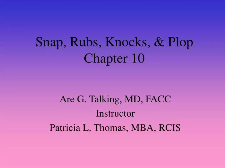
Snap, Rubs, Knocks, & Plop Chapter 10. Are G. Talking, MD, FACC Instructor Patricia L. Thomas, MBA, RCIS. Outline. Opening Snap of Mitral Stenosis Opening Snap of Tricuspid Stenosis Pericardial Friction Rub Pericardial Knock Mediastinal Crunch Tumor Plops.

E N D
Snap, Rubs, Knocks, & PlopChapter 10 Are G. Talking, MD, FACC Instructor Patricia L. Thomas, MBA, RCIS
Outline • Opening Snap of Mitral Stenosis • Opening Snap of Tricuspid Stenosis • Pericardial Friction Rub • Pericardial Knock • Mediastinal Crunch • Tumor Plops
Opening Snap of Mitral Stenosis • Heard in Early Diastole a characteristic sign of mitral stenosis • Sharp, medium frequency, & moderately loud sounds • Early diastole at the end of the isometric relaxation period • Occurs .06 to .12 sec after the A2 • Severe Stenosis-LA pressure is higher & forces the valve quickly forward to an early snap
Causes • In mitral stenosis, scarring & thickening of the valve leaflet edges has bound them together; the most common cause is rheumatic heart disease • Opening snap is a result of the anterior leaflet becoming taut, and snaps to a halt, producing a sharp early diastolic sound that is diagnostic of mitral stenosis
Where to Listen • Use the diaphragm of the stethoscope between the apex the lower left sternal border at the fourth intercostal space
Associated Findings • See figure 10-2 • Phonocardiogram of MS & Sinus Rhythm • Opening Snap crescendo diastolic murmur • Loud first heart sound • Accentuated presystolic component
Intensity of the Snap • Mitral Stenosis the opening snap is almost always heard • Moderately calcified valve leaflets • Soften opening snap • Severely calcified valve leaflets • No opening snap • Physical activity & opening snap • LA mean is high louder opening snap • Aortic insufficiency • May decrease the intensity of the OS because of the reduced rate of anterior motion of the mitral leaflets in early diastole
Pericardial Friction Rub • Inflammation of the pericardial membrane or pleural sac at the 3rd and 4th interspace at the left sternal border. Louder during inspiration • Scratchy, like sandpaper being used, a match being struck, or leather squeaking • Sound present when the epicardial & pericardial surfaces, roughened by inflammation, slide over one another during atrial & ventricular systole & during the passive motion of rapid ventricular filling • 3 components • Atrial Systolic (A) • Ventricular Systolic (V) • Ventricular Diastolic (D)
Percardial Knock • Sharp, high-pitched sound present in 90% or more of patients with constrictive pericarditis • Heard in diastole • Occurs .09 to .12 sec after S3 • See figure 10-5 • Occurs after Heart Surgery, radiation therapy, viral infection, TB pericarditis • Diaphragm of the stethoscope listen at the lower left sternal border
Tumor Plops • Clues to a Myxoma • Cardiac silhouette on X-Ray consistent with atrial enlargement • An ECG showing signs of LA enlargement • Light-headedness • A very short presystolic murmur • Extra sound in diastole
Tumor Plops • Left Atrial Tumors • Loud, low-frequency thud heard in early diastole & caused by abrupt movements of the tumor inside the LA • It strikes the wall of the chamber or comes to a sudden halt as the pedicle reaches the limit its stretch • Mitral Valve Vegetation • Echo demonstrates a physical movement of vegetation across the mitral valve apparatus • Listen with the diaphragm of the stethoscope at the point of PMI with patient in the left lateral position
THE ENDOFCHAPTER 10 Tilkian, Ara MD Understanding Heart Sounds and Murmurs, Fourth Edition, W.B. Sunders Company. 2002, pp. 107-120