Download

1 / 22
220 likes | 228 Views
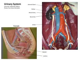
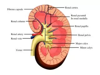
بسم الله الرحمن الرحيم Practical histopathology Lab 7 ( Medical Renal Biopsy ). Introduction. The urinary system , also known as the renal system , consists of the kidneys, ureters, bladder, and the urethra. Each kidney consists of millions of functional units called nephrons .

E N D
بسم الله الرحمن الرحيم Practical histopathology Lab 7 (Medical Renal Biopsy)
Introduction • The urinary system, also known as the renal system, consists of the kidneys, ureters, bladder, and the urethra. • Each kidney consists of millions of functional units called nephrons. • Nephron Consists of 2 parts : 1- Glomerulus (filtering system) tuft of capillaries surrounded by Bowman’s capsule 2- Tubules
Kidney disease diagnosis • Blood tests:measure the level of waste products, such as creatinine, urea and uric acid. • Urine tests:pH, Chemical Analyses (glucose, protein, bilirubin/urobilinogen & others) • Imaging tests: by using ultrasound to assess kidneys' structure and size.
4. Kidney biopsy remains the gold standard for the diagnosis and treatment of medical renal disease occurring in both native and transplanted kidneys. • In particular, kidney biopsy can be used to evaluate the type, extent, site, and nature of renal involvement in a disease. • Renal biopsy is a relatively low-risk procedure. • The pattern of light microscopic injury in the renal biopsy is typically the major clue to diagnosis.
Each biopsy is therefore evaluated for changes in glomeruli, tubules, interstitium, and vessels to arrive at a preliminary (and often final) diagnosis.
Functions of renal system • Removal of waste product from the body (mainly urea and uric acid). • Regulation of electrolyte balance (e.g. sodium, potassium and calcium). • Regulation of acid-base homeostasis. • Controlling blood volume and maintaining blood pressure.
Filtration • Inside each kidney are around a million tiny structures called nephronswhich filters blood to produce urine. • Arterioles in the kidneys deliver blood to a bundle of capillaries surrounded by a capsule called a glomerulus. • As blood flows through the glomerulus, much of the blood’s plasma is pushed out of the capillaries and into the capsule, leaving the blood cells and a small amount of plasma to continue flowing through the capillaries.
The liquid filtrate in the capsule flows through a series of tubules lined with filtering cells and surrounded by capillaries. • The cells surrounding the tubules selectively absorb water and substances from the filtrate in the tubule and return it to the blood in the capillaries. • At the same time, waste products present in the blood are secreted into the filtrate. • By the end of this process, the filtrate in the tubule has become urine containing only water, waste products, and excess ions. • The blood exiting the capillaries has reabsorbed all of the nutrients along with most of the water and ions that the body needs to function.
Storage and Excretion of Wastes • After urine has been produced by the kidneys, it is transported through the ureters to the urinary bladder. • The urinary bladder fills with urine and stores it until the body is ready for its excretion. • When the volume of the urinary bladder reaches anywhere from 150 to 400 milliliters, its walls begin to stretch and stretch receptors in its walls send signals to the brain and spinal cord.
These signals result in the relaxation of the involuntaryinternal urethral sphincter and the sensation of needing to urinate. • Urination may be delayed as long as the bladder does not exceed its maximum volume, but increasing nerve signals lead to greater discomfort and desire to urinate. • Urination is the process of releasing urine from the urinary bladder through the urethra and out of the body.
The process of urination begins when the muscles of the urethral sphincters relax, allowing urine to pass through the urethra. • At the same time that the sphincters relax, the smooth muscle in the walls of the urinary bladder contract to expel urine from the bladder.
Glomerulonephritis • Glomerulonephritis is inflammation of the tiny filters of the kidneys. • It occurs on its own or as part of another disease, such as lupus or diabetes. Severe or prolonged inflammation associated with glomerulonephritis can damage your kidneys. • Blood and protein in the urine are common problems that occur with glomerulonephritis. It can also result in kidney failure.
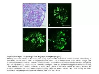
Causes 1- Infections • Post-streptococcal glomerulonephritis, Bacterial endocarditis, Viral infections). 2- Immune diseases • (Lupus, Goodpasture's syndrome, IgA nephropathy). 3- Conditions likely to cause scarring of the glomeruli • (High blood pressure, Diabetic kidney disease (diabetic nephropathy).
Polycystic kidney disease • A genetic condition resulting in large cysts in both kidneys that impair their function.
Papillary necrosis • Severe damage to the kidneys can cause chunks of kidney tissue to break off internally and clog the kidneys. • If untreated, the resulting damage can lead to total kidney failure.
Kidney cancer • It is not a single disease; it is made up of a number of different types of cancer that occur in this organ. • Each may have a distinct histologic type, have a different clinical course, respond differently to therapy, and be caused by alteration of a different gene. • It occurs in an inherited form and a sporadic, non-inherited form. • To identify the genes that cause sporadic, non-inherited cancer of the kidney, families with kidney cancer were studied to determine whether the genes that cause the inherited forms of renal cell carcinoma (RCC) might be involved in the development of the common forms of sporadic, non-inherited kidney cancer.
Although there have been remarkable advances in the development of immunologic forms of therapy for this disease, currently there is still no effective form of therapy for most patients with advanced renal carcinoma. • Those patients who present with advanced kidney cancer have a 2-year survival rate of <20%
Thanks a lot