Download
1 / 42
440 likes | 896 Views
S-114.740 Special Course in Communication and Cognition: Neural Plasticity. Iiro P. Jääskeläinen, Ph.D., Professor Cognitive Science and Technology Laboratory of Computational Engineering. What is plasticity?. Functional organization of the brain reflects adaptation to environment

E N D
S-114.740 Special Course in Communication and Cognition:Neural Plasticity Iiro P. Jääskeläinen, Ph.D., Professor Cognitive Science and Technology Laboratory of Computational Engineering
What is plasticity? • Functional organization of the brain reflects adaptation to environment • As long as the environment (and the neural systems) stay approximately the same, functional organization remains the same • Changes in the environment and in the neural systems (such as after a lesion) trigger plastic changes to facilitate re-adaptation
Different kinds of plasticity • Developmental plasticity (immature brain first begins to process sensory information) • Activity-dependent plasticity (changes in sensory input due to, e.g., eyesight problems) • Plasticity of learning and memory (e.g. discrimination training) • Injury-induced plasticity (following brain damage)
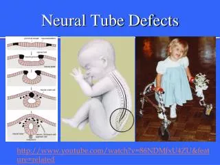
Development and plasticity • Critical sensitivity periods in development • Language acquisition (1st and 2nd) • Pruning as an underlying mechanism? • initially more connections than in the mature CNS • Damage early during development relatively minimal adverse effects (e.g., hydrocephalus findings)
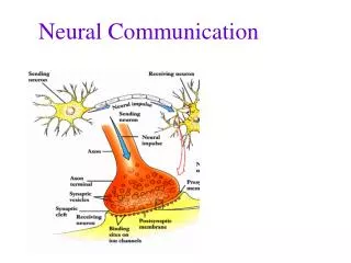
NMDA-receptors and synaptic plasticity • Convergent pre-synaptic activity leads to strenghtening of synaptic connections • Magnesium blockade of NMDA receptors is removed by depolarization Ca2+ influx plasticity
Cross-modal plasticity in congenitally deaf • These PET/MR images show increased neural activity in the superior temporal gyrus in congenitally deaf subjects when they viewed signs or sign-like movements, suggesting that auditory cortical regions may contribute to the processing of visual information in the deaf
Changes in sensory input induce plastic changes in somatotopy • Spinal cord injuries in adult monkeys result in somatosensory reorganization of the topographic map in area 3b. The region of the map that normally processes sensory information from the hand now receives sensory inputs from the face.
Plastic changes induced experimentally • Changing the external stimulus environment • Reversal of the visual world with goggles after a period of days, switching of the view to normal despite goggles • Sensory deprivation and hallucinations • Somatosensory two-point discrimination training changes in somatosensory homonculus
Short-term plasticity: a short definition • Influence of previous stimuli (i.e., memory), top-down effects (e.g., attention), and learning (longer-term plasticity), on how the sensory systems filter stimuli, enabling tracking of and reacting to relevant objects
Paired-pulse effects • The simplest form of short-term plasticity is perhaps manifested in paired-pulse effects • paired-pulse depression • paired-pulse facilitation • Short-lived changes in amplitude and latency of responses to the second stimulus of a pair • Sensory memory?
Neural tuning • Auditory-system neurons exhibit selective responses to certain stimulus attributes over others • Combined with PPD/PPF, neural tuning can explain short-term sensory memory
Differential adaptation of N1m(a) and N1m(p) explain the mismatch response Differential adaption of anterior and posterior sources contributing to the overall N1m response explains the differences in ECD loci between the MMNm and N1m Anterior N1m: slower in latency, sharp frequency tuning. Related to the ”what” processing stream? Posterior N1m: fast, only coarse frequency tuning. Related to the ”where” processing stream? Jääskeläinen et al. PNAS101: 6809–6814, 2004
Selective attention tunes responses to 3-D vs. phonetic Stimulus pairs varying in both phonetic (/ö/ vs. /ä/) and 3-D location features Task of the subject: is the pair same or different with respect to the preceding pair in 3-D location or phonetic content? Passive ”ignore” condition Ahveninen, Jääskeläinen et al. in preparation
Combined 3-T fMRI (Siemens Trio) and 306-channel MEG (Neuromag VectorView) data suggested sharper neural tuning in areas posterior to primary auditory cortex. Selective attention to 3-D significantly augmented this.
Corroborating macaque findings on the ”what” and ”where” • Monkey studies suggesting anterior (AL) ”what” and posterior (CL) ”where” processing pathways in the auditory cortex • Spatial location vs. species-specific vocalizations • Visual system analogy? Rauschecker & Tian PNAS 97: 11800–11806, 2000
Gain vs. tuning: an open question • Several studies have contrasted the hypotheses of gain vs. tuning as the neural basis for selective attention • Possible tuning mechanisms include narrowing of and shifts in tuning curve
Is there tonotopy at all? • While BFs to pure tones disclose tonotopic organization, the responses even at BF are not vigorous • Stimulation sweeping at certain speed over the BF elicit most robust responses in AC neurons • Spectrotemporal receptive fields
Dynamic STRF changes in AC Fritz J et al. Nature Neuroscience 6:1217-1223, 2003
Modulation of primary auditory cortex activity by visual speech During continous scanner noise, seeing movies of visual articulations vs. a still-face baseline significantly activated the human primary auditory cortex Dynamic modulation of primary auditory cortex STRFs aiding speech perception? Pekkola et al. submitted
AC vs. subcortical structures • Corticofugal influence: electrical stimulation of auditory cortex causes modulation of STRFs at lower auditory system structures, MGB, IC, even cochlea! • Animal data suggest that the lower one goes, the longer time it takes to see such changes • AC as the ”initiator” of modulatory effects?
Short-term plasticity and the somatosensory system • Local anesthesia of a finger causes relatively rapid changes in cortical representation areas • These changes are quickly reversed to normal upon normalization of stimulation • ”Dormant” connections between areas as underlying neural mechanism?
Attention and gain in somatosensation • When attention is directed to the tactile stimulus, the response of the neurons in the somatosensory cortex is enhanced, compared to when attention is directed to visual stimuli.
Attention and plastic changes • Attention to certain stimulus features required for short-term plastic changes to occur • Transfer of short-term plastic changes to long-term ones?
Neurochemistry and plasticity • Selective lesions of central noradrenergic pathways impair recovery after a subsequent injury to the cerebral cortex. Drugs that deplete central norepinephrine, block alpha 1-adrenergic receptors, or decrease norepinephrine release (alpha 2-adrenergic receptor agonists) impede recovery whereas drugs that increase norepinephrine release (alpha 2-adrenergic receptor antagonists) or sympathomimetics (amphetamine) facilitate recovery • N.B. NE is a neurochemical correlate of attention! • Also, acetylcholine suggested to be vital for plasticity
Brain injury, rehabilitation and recovery • How quickly does the injury occur? • Brain tumors, hydrocephalus slow destruction of brain matter, time for adaptive / plastic changes • Brain tumors can be large before any symptoms are noticed • Stroke: sudden loss of areas, drastic behavioral / cognitive effects
”Spontaneous” recovery • ”Spontaneous” recovery from, e.g., stroke • Quick recovery of functions during the first three months after injury • Slower recovery thereafter
Re-occurrence of injury • After having sufffered brain damage (e.g., stroke), another stroke usually has significantly larger detrimental effects • ”Plastic reserve has been drained”
Rehabilitation • Circumventing the problem • anterograde amnesia after stroke: learning to use notebook • relatively effortless way to correct problem • Rehabilitation of function • anterograde amnesia after stroke: performing highly specific memory tasks, thus enhancing memory performance • diagnostics problems, persistence
Rehabilitation • Needs to be specific • loss of visual field (scotoma) • attention to to stimulation at the edges of the scotoma result gradually in smaller scotoma size • attention required! • How to design specific rehabilitation of e.g. executive functions? • Symptom self-recognition low
Imaginary training in rehabilitation? • Paralysis due to stroke may prevent early participation in a rehabilitation program • Similar network of cerebral structures (e.g., premotor cortex) is activated when normal control subjects execute physically or imagine a sequence of up-down foot movements mental practice with motor imagery can be used as a therapeutic approach to keep active the neural circuits involved in locomotion, facilitating the rehabilitation of patients who sustained damage to the brain(?)
Neurogenesis in the brain • Traditionally thought that new neurons are not produced in the brain • Recent studies have yielded tentative evidence for neurogenesis in, for instance, hippocampus even in adult brain • N.B. glia form impenetrable scars after brain injury • Also, methods are being developed wherein ”stem cells” are injected to brain that develop into neurons
Stem cells • Adult stem cells exists in the brain in small numbers, remaining quiescent (non-dividing) for many years until activated by e.g. disease / tissue injury. • Effort to find ways to grow adult stem cells in cell culture and manipulate them to generate specific cell types so they can be used to treat injury or disease. Some examples of potential treatments include replacing the dopamine-producing cells in the brains of Parkinson's patients.