Download
1 / 30
370 likes | 915 Views
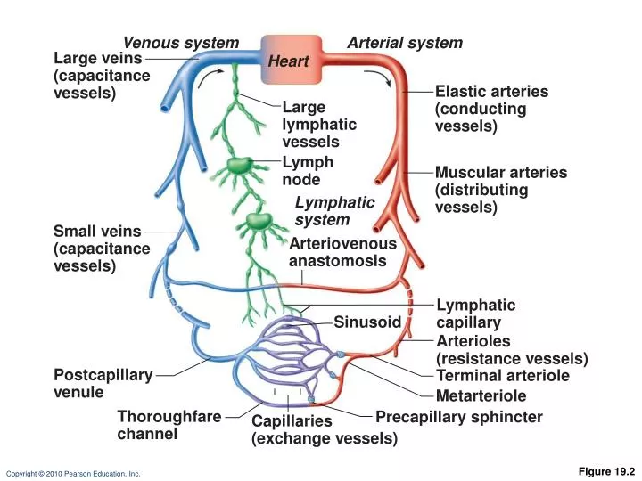
Arterial system. Venous system. Large veins (capacitance vessels). Heart. Elastic arteries (conducting vessels). Large lymphatic vessels. Lymph node. Muscular arteries (distributing vessels). Lymphatic system. Small veins (capacitance vessels). Arteriovenous anastomosis.

E N D
Arterial system Venous system Large veins (capacitance vessels) Heart Elastic arteries (conducting vessels) Large lymphatic vessels Lymph node Muscular arteries (distributing vessels) Lymphatic system Small veins (capacitance vessels) Arteriovenous anastomosis Lymphatic capillary Sinusoid Arterioles (resistance vessels) Postcapillary venule Terminal arteriole Metarteriole Thoroughfare channel Precapillary sphincter Capillaries (exchange vessels) Figure 19.2
Tunica intima Valve • Endothelium • Subendothelial layer Internal elastic lamina Tunica media (smooth muscle and elastic fibers) External elastic lamina Tunica externa (collagen fibers) Lumen Vein Lumen Artery Capillary network Basement membrane Endothelial cells Capillary (b) Figure 19.1b
Pericyte Red blood cell in lumen Intercellular cleft Endothelial cell Basement membrane Tight junction Pinocytotic vesicles Endothelial nucleus (a) Continuous capillary. Least permeable, and most common (e.g., skin, muscle). Figure 19.3a
Pinocytotic vesicles Red blood cell in lumen Fenestrations (pores) Endothelial nucleus Intercellular cleft Basement membrane Endothelial cell Tight junction (b) Fenestrated capillary. Large fenestrations (pores) increase permeability. Occurs in special locations (e.g., kidney, small intestine). Figure 19.3b
Endothelial cell Red blood cell in lumen Large intercellular cleft Tight junction Nucleus of endothelial cell Incomplete basement membrane (c) Sinusoidal capillary. Most permeable. Occurs in special locations (e.g., liver, bone marrow, spleen). Figure 19.3c
Vascular shunt Precapillary sphincters Thoroughfare channel Metarteriole True capillaries Terminal arteriole Postcapillary venule (a) Sphincters open—blood flows through true capillaries. Terminal arteriole Postcapillary venule (b) Sphincters closed—blood flows through metarteriole thoroughfare channel and bypasses true capillaries. Figure 19.4
Vein Artery (a) Figure 19.1a
Pulmonary blood vessels 12% Systemic arteries and arterioles 15% Heart 8% Capillaries 5% Systemic veins and venules 60% Figure 19.5
Physiology of Circulation: Definition of Terms • Blood pressure (BP) • Force per unit area exerted on the wall of a blood vessel by the blood • Expressed in mm Hg • Measured as systemic arterial BP in large arteries near the heart • The pressure gradient provides the driving force that keeps blood moving from higher to lower pressure areas
Physiology of Circulation: Definition of Terms • Peripheral resistance • Opposition to flow • Measure of amount of friction blood encounters • Generally encountered in peripheral circulation • Three important sources of resistance • Blood viscosity • Total blood vessel length • Blood vessel diameter
Arterial Blood Pressure • Systolic pressure: pressure exerted during ventricular contraction • Diastolic pressure: lowest level of arterial pressure • Pulse pressure = difference between systolic and diastolic pressure
Factors Aiding Venous Return • Respiratory “pump”: pressure changes created during breathing move blood toward the heart by squeezing abdominal veins as thoracic veins expand • Muscular “pump”: contraction of skeletal muscles “milk” blood toward the heart and valves prevent backflow • Vasoconstriction of veins under sympathetic control
Valve (open) Contracted skeletal muscle Valve (closed) Vein Direction of blood flow Figure 19.7
Maintaining Blood Pressure • The main factors influencing blood pressure: • Cardiac output (CO) • Peripheral resistance (PR) • Blood volume
Exercise BP activates cardiac centers in medulla Parasympathetic activity Activity of respiratory pump (ventral body cavity pressure) Sympathetic activity Activity of muscular pump (skeletal muscles) Epinephrine in blood Sympathetic venoconstriction Venous return Contractility of cardiac muscle EDV ESV Stroke volume (SV) Heart rate (HR) Initial stimulus Physiological response Cardiac output (CO = SV x HR Result Figure 19.8
3 Impulses from baroreceptors stimulate cardioinhibitory center (and inhibit cardioacceleratory center) and inhibit vasomotor center. 4a Sympathetic impulses to heart cause HR, contractility, and CO. 2 Baroreceptors in carotid sinuses and aortic arch are stimulated. 4b Rate of vasomotor impulses allows vasodilation, causing R 5 CO and R return blood pressure to homeostatic range. 1 Stimulus: Blood pressure (arterial blood pressure rises above normal range). Homeostasis: Blood pressure in normal range 1 Stimulus: Blood pressure (arterial blood pressure falls below normal range). 5 CO and R return blood pressure to homeostatic range. 4b Vasomotor fibers stimulate vasoconstriction, causing R 2 Baroreceptors in carotid sinuses and aortic arch are inhibited. 4a Sympathetic impulses to heart causeHR, contractility, and CO. 3 Impulses from baroreceptors stimulate cardioacceleratory center (and inhibit cardioinhibitory center) and stimulate vasomotor center. Figure 19.9
Arterial pressure Indirect renal mechanism (hormonal) Direct renal mechanism Baroreceptors Sympathetic stimulation promotes renin release Kidney Renin release catalyzes cascade, resulting in formation of Angiotensin II Aldosterone secretion by adrenal cortex ADH release by posterior pituitary Filtration Sodium reabsorption by kidneys Water reabsorption by kidneys Vasoconstriction ( diameter of blood vessels) Blood volume Initial stimulus Physiological response Arterial pressure Result Figure 19.10
Fluid loss from hemorrhage, excessive sweating Crisis stressors: exercise, trauma, body temperature Bloodborne chemicals: epinephrine, NE, ADH, angiotensin II; ANP release Activity of muscular pump and respiratory pump Release of ANP Dehydration, high hematocrit Body size Conservation of Na+and water by kidney Blood volume Blood pressure Blood pH, O2, CO2 Blood volume Baroreceptors Chemoreceptors Venous return Activation of vasomotor and cardiac acceleration centers in brain stem Diameter of blood vessels Blood viscosity Blood vessel length Stroke volume Heart rate Cardiac output Peripheral resistance Initial stimulus Physiological response Result Mean systemic arterial blood pressure Figure 19.11
Blood Flow Through Body Tissues • Blood flow (tissue perfusion) is involved in • Delivery of O2 and nutrients to, and removal of wastes from, tissue cells • Gas exchange (lungs) • Absorption of nutrients (digestive tract) • Urine formation (kidneys) • Rate of flow is precisely the right amount to provide for proper function
Brain Heart Skeletal muscles Skin Kidney Abdomen Other Total blood flow at rest 5800 ml/min Total blood flow during strenuous exercise 17,500 ml/min Figure 19.13
Metabolic Controls • Vasodilation of arterioles and relaxation of precapillary sphincters occur in response to • Declining tissue O2 • Substances from metabolically active tissues (H+, K+, adenosine, and prostaglandins) and inflammatory chemicals
Metabolic Controls • Effects • NO is the major factor causing vasodilation • Vasoconstriction is due to sympathetic stimulation and endothelins
Myogenic Controls • Myogenic responses of vascular smooth muscle keep tissue perfusion constant despite most fluctuations in systemic pressure • Passive stretch (increased intravascular pressure) promotes increased tone and vasoconstriction • Reduced stretch promotes vasodilation and increases blood flow to the tissue
Intrinsic mechanisms (autoregulation) Extrinsic mechanisms • Maintain mean arterial pressure (MAP) • Redistribute blood during exercise and thermoregulation • Distribute blood flow to individual organs and tissues as needed Amounts of: Nerves Sympathetic pH O2 a Receptors Epinephrine, norepinephrine Metabolic controls b Receptors Amounts of: CO2 Angiotensin II K+ Hormones Prostaglandins Antidiuretic hormone (ADH) Adenosine Nitric oxide Atrial natriuretic peptide (ANP) Endothelins Myogenic controls Stretch Dilates Constricts Figure 19.15
Long-Term Autoregulation • Angiogenesis • Occurs when short-term autoregulation cannot meet tissue nutrient requirements • The number of vessels to a region increases and existing vessels enlarge • Common in the heart when a coronary vessel is occluded, or throughout the body in people in high-altitude areas
Blood Flow: Skeletal Muscles • At rest, myogenic and general neural mechanisms predominate • During muscle activity • Blood flow increases in direct proportion to the metabolic activity (active or exercise hyperemia) • Local controls override sympathetic vasoconstriction • Muscle blood flow can increase 10 or more during physical activity
Blood Flow: Skin • Blood flow to venous plexuses below the skin surface • Varies from 50 ml/min to 2500 ml/min, depending on body temperature • Is controlled by sympathetic nervous system reflexes initiated by temperature receptors and the central nervous system
HP =hydrostatic pressure • Due to fluid pressing against a wall • “Pushes” • In capillary (HPc) • Pushes fluid out of capillary • 35 mm Hg at arterial end and 17 mmHg at venous end of capillaryin this example •In interstitial fluid (HPif) • Pushes fluid into capillary • 0 mm Hg in this example Arteriole Venule Interstitial fluid Capillary Net HP—Net OP (17—0)—(26—1) Net HP—Net OP (35—0)—(26—1) OP =osmotic pressure • Due to presence of nondiffusible solutes (e.g., plasma proteins) • “Sucks” •In capillary (OPc) • Pulls fluid into capillary • 26 mm Hg in this example •In interstitial fluid (OPif) • Pulls fluid out of capillary • 1 mm Hg in this example Net HP 35 mm Net OP 25 mm Net OP 25 mm Net HP 17 mm NFP (net filtration pressure) is 10 mm Hg; fluid moves out NFP is ~8 mm Hg; fluid moves in Figure 19.17
Initial stimulus Acute bleeding (or other events that cause blood volume loss) leads to: Physiological response 1. Inadequate tissue perfusion resulting in O2 and nutrients to cells 2. Anaerobic metabolism by cells, so lactic acid accumulates 3. Movement of interstitial fluid into blood, so tissues dehydrate Signs and symptoms Result Baroreceptor firing reduced (by blood volume and pressure) Hypothalamus activated (by pH and blood pressure) Chemoreceptors activated (by in blood pH) Brain Major effect Minor effect Neurons depressed by pH Activation of respiratory centers Cardioacceleratory and vasomotor centers activated Sympathetic nervous system activated ADH released Intense vasoconstriction (only heart and brain spared) Heart rate Central nervous system depressed Kidney Renal blood flow Renin released Adrenal cortex Angiotensin II produced in blood Aldosterone released Kidneys retain salt and water Water retention Rate and depth of breathing Tachycardia, weak, thready pulse Skin becomes cold, clammy, and cyanotic Restlessness (early sign) Coma (late sign) Urine output Thirst Blood pressure maintained; if fluid volume continues to decrease, BP ultimately drops. BP is a late sign. CO2blown off; blood pH rises Figure 19.18