Download
1 / 10
110 likes | 209 Views
Title: Co-delivery of antigen and IL-12 by Venezuelan equine encephalitis virus replicon vectors enhances antigen-specific immune responses and anti-tumor effects. Journal : Cancer Immunology, Immunotherapy

E N D
Title: Co-delivery of antigen and IL-12 by Venezuelan equine encephalitis virus replicon vectors enhances antigen-specific immune responses and anti-tumor effects. Journal: Cancer Immunology, Immunotherapy Authors: Takuya Osada1, Peter Berglund5, Michael A. Morse2,4, Bolyn Hubby5, Whitney Lewis6, Donna Niedzwiecki4, Xiao Yi Yang1, Amy Hobeika1, Bruce Burnett4, Gayathri R. Devi1,4, Timothy M. Clay1,3,4,, Jonathan Smith5 , and H. Kim Lyerly1,3,4. Affiliation: Duke University Medical Center, Departments of 1Surgery, 2Medicine, 3Immunology, and 4Duke Comprehensive Cancer Center, Durham, NC 27710, U.S.A. 5Liquidia Technologies, RTP, NC 27709, 6Precision Biosciences, Durham, NC 27701, U.S.A Corresponding Author: H. Kim Lyerly, MD Duke University Medical Center Department of Surgery Box 2606 MSRB1 Rm 433b Research Dr Durham, NC 27710 Lyerl001@mc.duke.edu Tel: 919-684-5613
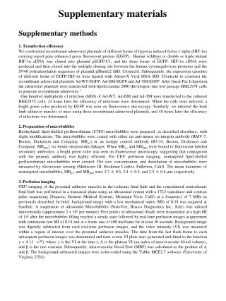
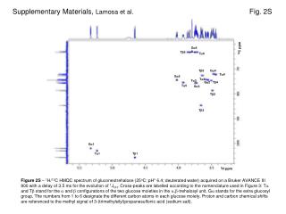
Supplementary Materials Immunofluorescent staining of VRP-CEA(6D) infected Vero cells Vero cells, grown to near confluency, were infected by removing the culture medium and overlaying the cell monolayer with medium containing VRP-CEA(6D) or control HIV-gag VRP (moi 100), incubated overnight. Productive infection was determined by staining for CEA expression under permeabilized and non-permeabilized conditions followed by visualization by immunofluorescence. After fixation of cells with 4% paraformaldehyde for 10 min, permeabilization was performed with 0.01% triton x-100 in PBS for 7 min at room temperature. Cells were labeled with mouse anti-CEA monoclonal antibody (Invitrogen, Carlsbad, CA) for 60 min, then with FITC-conjugated anti-mouse IgG secondary antibody for 45 min. Similarly, IL-12 p70 expression by VRP-IL-12 infected Vero cells was verified using immunofluorescence staining (data not shown). Western blot analysis of cell lysate from VRP-infected Vero cells. Cell lysates were made from VRP-CEA(6D)-infected Vero cells, To confirm expression of full length CEA and normal glycosylation of the molecule, a half of the cell lysate was incubated with/without Peptide N Glycosidase F (PNGase F, 500 units/µg protein, Sigma) for 3 h at 37°C to deglycosylate CEA. Similarly, Vero cells were infected with VRP-IL-12 (moi 100), incubated for 16 h, and cell lysate was made and processed for SDS-PAGE. Western blot analysis was performed using an anti-mouse IL-12 antibody (Invitrogen). Recombinant murine IL-12 (rIL-12) was loaded at various amounts (40, 80, 100, 200, 400 ng protein/lane) to compare the separation of the p40 and p35 subunits in the denaturing gel with VRP-expressed monomeric fusion polypeptide.
A B CEA replicon GAG replicon permeabilized non- permeabilized A B C C D Lane: 1 2 3 4 CEA repliconRNA: + - + - PNGase: + + - - Supplementary Figure 1
Supplementary Figure 1.CEA expression by VRP-CEA(6D) infected cells. (A) VRP-CEA vector construct. (B) Vero cells, grown to near confluency, were infected by removing the culture medium and overlaying the cell monolayer with medium containing VRP-CEA(6D) or control HIV-gag VRP, incubated overnight. Productive infection was determined by staining for CEA expression under permeabilized and non-permeabilized conditions followed by visualization by immunofluorescence (IFA). As demonstrated, CEA staining was localized intracellularly as well as on the plasma membrane in cells infected with VRP-CEA(6D). The non-cytoplasmic, reticular pattern of the intracellular signal was consistent with biosynthesis through ER-Golgi for secretion on transmembrane insertion. (C) Western blot was used to confirm the expression of full length CEA in lysates from VRP-CEA(6D) infected Vero cells.We confirmed that full length CEA protein was expressed from the CEA replicon and yielded a shorter fragment consistent with the putative 76.8 kDa molecular weight when deglycosylated. These data demonstrate that CEA is appropriately translocated and post-translationally modified, and transported through the Golgi to the plasma membrane.
A B Supplementary Figure 2
Supplementary Figure 2. IL-12 production by VRP-IL-12 infected cells. (A) VRP-IL12 vector construct. The vector was designed to express murine IL-12 in the form of a single polypeptide where the p35 and p40 subunits are fused into one open reading frame separated by a short linker sequence derived from the elastin gene. (B) Functional p70 expression was verified using Western blot on cell lysates from Vero cells infected with VRP-IL12. At 16 h post infection, cells were lysed and processed for SDS-PAGE, and Western blot analysis using an IL-12 specific antibody. Cell lysate (pERK-IL12) was loaded in the right-most lane and recombinant murine IL-12 (rIL-12) was loaded at various amounts (40, 80, 100, 200, 400 ng/lane), as indicated. rIL-12 migrates as two bands, corresponding to the separation of the p40 and p35 subunits in this denaturing gel, whereas the VRP-expressed monomeric fusion polypeptide appears as a 70 kDa band. Expected migration patterns of the polypeptides are indicated by the arrows.
CD80 CD86 73.2 79.8 Mock 87.8 453.6 VRP-IL12 (moi 100) 553.7 87.1 VRP-Empty 185.7 76.3 LPS Supplementary Figure 3
Supplementary Figure 3. VRP-IL-12 infection of DC leads to maturation of DC. Maturation status of VRP-IL-12 infected DC (MOI 10, 100) was analyzed after 24 hours of infection. Cells were stained with FITC-labeled anti-mouse CD14, APC-labeled anti-I-A/I-E, and PE-lebeled anti-CD80 or anti-CD86 mAb. Blocking of Fcg receptor with anti-mouse CD16/CD32 was made before staining. CD14-negative/MHC class II-positive DCs were analyzed for CD80/CD86 expression. Solid histograms show staining with PE-labeled anti-CD80 or anti-CD86 mAb, and open histograms show control staining with PE-labeled control IgG. Value of Mean Fluorescence Intensity is shown in each histogram.
* * CEA Antibody Response CEA Cellular Response SFC/1e6 lymphocytes Reciprocal Titer CEA protein CEA protein CEA protein + VRP-IL12 CEA protein + VRP-IL12 Supplementary Figure 4
Supplementary Figure 4. Co-administration ofVRP-IL-12 enhanced cellular and humoral immune response against CEA with CEA protein vaccination. To assess the immunomodulatory effect of VRP-IL-12 for a different vaccine platform, mice were immunized with carcinoembryonic antigen (CEA(6D)) protein with/without co-injection of VRP-IL-12 (5x10⁵ I.U). Immunization was repeated twice, 3 weeks apart. Immune monitoring assays were performed 7 days after the second immunization. Anti-CEA immune responses were analyzed by IFN-gamma ELISPOT assay or anti-CEA ELISA. VRP-IL-12 significantly enhanced anti-CEA cellular and humoral immune responses induced by CEA(6D) protein vaccine. *p<0.01


![[Supplementary materials]](https://cdn1.slideserve.com/2110594/slide1-dt.jpg)