Download
1 / 28
280 likes | 336 Views
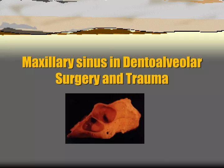
Maxillary sinus in Dentoalveolar Surgery and Trauma. Oro-antral fistula:. Invasion of the maxillary sinus and establishment of a direct communication with the oral cavity is referred to as an oro-antral fistula. Fistula:.

E N D
Oro-antral fistula: Invasion of the maxillary sinus and establishment of a direct communication with the oral cavity is referred to as an oro-antral fistula.
Fistula: • Is a biological tract that connect an anatomical cavity with the external surfaces or another anatomical cavity, (unlike sinus tract). It is always lined with a stratified squamous epithelium and the potency of the tract is preserved until epithelial cells scraped off.
Factors influencing creation of oro-antral fistula: • Teeth size and configuration of the roots. • Hypercementosis and bulbous roots. • Density of alveolar bone and thickness of sinus floor • Size of the sinus. • Relation of sinus to the root of upper teeth. • Rough extraction and misguided manipulation. • Apical pathosis and attached granulomas. • Periodontal diseases which may erode sinus floor. • Presence of cysts and neoplasm. • Invasive surgery e.g. cleft and dental implants placement.
Signs and symptoms of newly created oro-antral fistula: • Antral floor attached to roots apices of extracted tooth or teeth. • Fracture of the alveolar process or the tuberosity. • Evidence of air stream passing from nostril. • Bubbling of blood from the socket or nostril. • Change in speech tone and resonance. • Radiographical evidence of sinus involvement.
Confirmation of existence oforo-antral fistula • Instruct patient to occlude the nostrils and blow genteelly “nose-blowing’ test”. • If nose-blowing’ test is negative, don’t explore the opening with suction tip and/or probes. • Don’t attempt to irrigate the sinus to confirm diagnosis, especially if the sinus drainage is impaired due to pre-existed sinusitis. • Always check radiograph for the continuity of sinus floor and presence of entrapped foreign body.
Displacement of tooth or root into the maxillary sinus lining or the sinus cavity proper • It is basically a mishap incident results from a neglected act by the operator while applying wrong force. • Occurs rarely but the 3rd molar and 2nd premolar are the most at risk of dislodgment. • May occur during forceful mouth opening of unconscious patient when using mouth gag of periodontaly involved teeth. • May occur with severe maxillofacial injures. • In association with poor surgical technique.
Immediate management/ investigations • Confirm the existence of oro-antral fistula and the presence of tooth or root in sinus using dental,occlusal, panoramic and occipito- mental radiographs. • Locate the precise position of the foreign body within the sinus lining or in the sinus cavity proper “head-shaking test”.
Immediate management/ foreign body retrieval • Reflect mucoperiosteal flap. • Reduce alveolar bone height. • Retrieve the tooth or the root by permitting their movement away from the sinus. • If root or tooth dislodged into the sinus proper, consider Caldwell-luc approach. • Undermine the flap and replace across the bony defect.
Immediate management/closure of the defect • Relieve the tension of the flap by serving the periostium. • Advance the flap across the defect and beyond. • Anchor the corner of the flap and approximate the edges using horizontal mattress sutures.
Alternative method of immediate repair of oro-antral fistulabecomes less popular due to transmission of infection;BSE-FJD • Use of lyophilized sterilized collagen sheet: reflect mucoperiosteal flap. reduce the height of bony socket . trim the collagen sheet to cover only the bony defect. slide underneath buccal and palatal extensions of the flap. secure the graft by suturing the flap extensions.
Postoperative care/ Home car • Acrylic base plate (surgical stint) may be prescribed to add additional support to the area. • Patient should avoid forceful nasal blowing, if forced to do so, no occluding of nares. • Oral hygiene must be kept optimum.
Postoperative care/ medications • Antibiotic e.g. Penicillin or penicillin derivatives • Analgesic and NSAI e.g. Paracetamol, profen (PRN) • Nasal decongestant e.g. Ephedrine or otrivin nasal drops 3 drops/ 3times daily / 7 days • Steam inhalation e.g. menthol and benzoin 40 good sniffs should follows nasal drops
Precaution measures in prevention of oro-antral fistula • Don’t apply forceps to maxillary posterior teeth unless enough tooth structure is sufficient to permit the blades to be applied. • Fractured root apex, in particular the palatal root of vital maxillary molar is better to put on probation. • Removal of isolated maxillary molar or extraction in a patient with H/O antral involvement must warrant careful radiographical assessment. • Removal of any maxillary root, if indicated, should be preceded by accurate localization via trans-alveolar approach. • Surgeon must provide a support for blood clot to organize by means of figure eight suture or using of surgical stint.
Chronic oro-antral fistula/persistent oro-antral communication It might be a complication of: • Unrecognized (overlooked) fistula. • Untreated fistula. • Failure of spontaneous closure of OAF. • Failure of surgically repaired fistula
Signs and symptoms of chronic fistula • Reflux of food and drinks. • Loss of denture stability. • Intermittent episode of pain and local tenderness. • Foul-tasting discharge. • Sings and symptoms of chronic sinusitis.
Primarily management of chronic OAF it is aimed to eliminate any sinus infection: • Excision of any mucosal polyp or purulent granulation to promote drainage. • Regular irrigation with warm water or saline. • Single course of antibiotics and nasal inhalation and decongestant. • Acrylic base plate.
Surgical management/Principles and requirements • Success of operation is not always garneted. • Flap should have good blood supply. • Flap tissue must be handled genteelly. • Flap should lie in its new position without tension. • Good haemostasis must be achieved before discharging the patients.
Surgical management/types of repair • Buccal advancement flap
Surgical management/types of repair • Bridge (pedicle) flap
Surgical management/types of repair • Palatal transposition flap
Surgical management/types of repair • Rotation palatal flap This is only possible in edentulous patients; exclusively indicated for edentulous patient.
Exploration of maxillary sinuous/Caldwell-luc approach • Recovery of entrapped foreign body from the sinus cavity proper; displaced tooth or root. • Excision of sinus polyps,tumors and cysts. • Treatment of blow out orbital fracture. • Grafting of maxillary sinus.
Fracture of maxillary tuberosity/predisposing factors • Expansion of sinus deep into the tuberosity. • Maxillary molar teeth of divergent or hypercementosed roots. • Maxillary tooth geminated or pathologically fused with adjacent one. • Over-eruption of isolated maxillary tooth. • Existence of pathological lesion. • Increase in bone density and fragility.
Management of tuberosity fracture • In the event of tuberosity fracture: Forceps extraction is to be abandoned. Surgical extraction then to be instituted. Dissection of bony fragment with attached tooth. Approximation of flap using mattress suturing technique.
Alternatively,In case of large scale fracture of the tuberocity and alveolar bone • bony fragment may be splinted in-situ using any method of fixation; Wiring or plating • and tooth extraction is to be delayed until union occurs.
EXTRA TIPS…….. BEFORE THE END OF THIS YEARMalignant disease of maxilla and maxillary sinus/Sings and symptoms • None dental maxillary pain • None inflammatory swelling of cheek • Loss of teeth • Epistaxis and gingival bleeding • Narrowing of the palpebral fissure • Depression of the corner of the mouth • Intra-oral swelling obliterated the sulcus • Proptosis and facial parasthesia and numbness • Radiographical evidence of invasive tumor