Download
1 / 57
580 likes | 587 Views
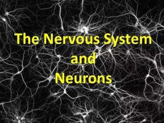
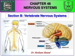
CHAPTER 44 Neurons and Nervous Systems. Chapter 44: Neurons and Nervous Systems. Nervous Systems: Cells and Functions Neurons: Generating and Conducting Nerve Impulses Neurons, Synapses, and Communication Neurons in Networks. Nervous Systems: Cells and Functions.

E N D
Chapter 44: Neurons and Nervous Systems Nervous Systems: Cells and Functions Neurons: Generating and Conducting Nerve Impulses Neurons, Synapses, and Communication Neurons in Networks
Nervous Systems: Cells and Functions • Nervous systems consist of cells that process and transmit information. 3
Nervous Systems: Cells and Functions • Sensory cells transduce information from the environment and body. • This communicates commands to effectors such as muscles or glands. 4
Nervous Systems: Cells and Functions • The nervous systems of different species vary, but all are composed of cells called neurons. Review Figures 44.1, 44.2 5
figure 44-01.jpg Figure 44.1 Figure 44.1
Nervous Systems: Cells and Functions • In vertebrates, brain and spinal cord form the central nervous system. • They communicate with other body tissues via the peripheral nervous system. 8
figure 44-02.jpg Figure 44.2 Figure 44.2
Nervous Systems: Cells and Functions • Neurons receive information mostly via their dendrites and transmit information over their axons. • They function in networks. Review Figure 44.3 9
figure 44-03a.jpg Figure 44.3 – Part 1 Figure 44.3 – Part 1
figure 44-03b.jpg Figure 44.3 – Part 2 Figure 44.3 – Part 2
Nervous Systems: Cells and Functions • Information that neurons process is in the form of electrical events in their plasma membranes. • Where neurons and other cells meet, information is transmitted mostly by release of chemical signals called neurotransmitters. 12
Nervous Systems: Cells and Functions • Glial cells physically support neurons and perform many housekeeping functions. • Schwann cells and oligodendrocytes produce myelin, which insulates neurons. • Astrocytes create the blood–brain barrier. Review Figure 44.4 13
figure 44-04.jpg Figure 44.4 Figure 44.4
Neurons: Generating and Conducting Nerve Impulses • Neurons have an electric charge difference across their plasma membranes. • This resting potential is created by ion pumps and channels. Review Figure 44.5 15
figure 44-05.jpg Figure 44.5 Figure 44.5
Neurons: Generating and Conducting Nerve Impulses • The sodium–potassium pump concentrates K+ ions on the insides and Na+ ions on the outsides of neurons. • Ion channels allow K+ ions to leak out, leaving behind unbalanced negative charges, leading to the resting potential. Review Figures 44.6, 44.7 17
figure 44-06.jpg Figure 44.6 Figure 44.6
figure 44-07.jpg Figure 44.7 Figure 44.7
Neurons: Generating and Conducting Nerve Impulses • A potassium equilibrium potential exists when an electric charge that develops across the membrane is sufficient to prevent net diffusion of potassium ions down their concentration gradient. • This potential can be calculated with the Nernst equation. Review Figure 44.8 20
figure 44-08.jpg Figure 44.8 Figure 44.8
Neurons: Generating and Conducting Nerve Impulses • The resting potential is perturbed when ion channels open or close, thus changing plasma membrane permeability to charged ions. • Thus, neurons become depolarized or hyperpolarized in response to stimuli. Review Figure 44.9 22
figure 44-09a.jpg Figure 44.9 – Part 1 Figure 44.9 – Part 1
figure 44-09b.jpg Figure 44.9 – Part 2 Figure 44.9 – Part 2
Neurons: Generating and Conducting Nerve Impulses • Rapid reversals in charge across portions of the plasma membrane, resulting from opening and closing of voltage-gated sodium and potassium channels, produce action potentials. • These changes occur when the plasma membrane depolarizes to a threshold level. Review Figure 44.10 25
figure 44-10.jpg Figure 44.10 Figure 44.10
Neurons: Generating and Conducting Nerve Impulses • Action potentials are conducted down axons because of local current flow • This depolarizes adjacent regions of membrane and brings them to threshold for the opening of voltage-gated sodium channels. Review Figure 44.11 27
figure 44-11a.jpg Figure 44.11 – Part 1 Figure 44.11 – Part 1
figure 44-11b.jpg Figure 44.11 – Part 2 Figure 44.11 – Part 2
figure 44-11c.jpg Figure 44.11 – Part 3 Figure 44.11 – Part 3
Neurons: Generating and Conducting Nerve Impulses • Patch clamping allows us to study single ion channels. Review Figure 44.12 31
figure 44-12.jpg Figure 44.12 Figure 44.12
Neurons: Generating and Conducting Nerve Impulses • In myelinated axons, the action potentials appear to jump between nodes of Ranvier, patches of plasma membrane not covered by myelin. Review Figure 44.13 33
figure 44-13a.jpg Figure 44.13 – Part 1 Figure 44.13 – Part 1
figure 44-13b.jpg Figure 44.13 – Part 2 Figure 44.13 – Part 2
Neurons, Synapses, and Communication • Neurons communicate with each other and other cells at specialized junctions called synapses, where plasma membranes of two cells come close together. 36
Neurons, Synapses, and Communication • The classic chemical synapse is the neuromuscular junction, a synapse between a motor neuron and muscle cell. • Its neurotransmitter is acetylcholine, which causes a depolarization of the postsynaptic membrane when it binds to its receptor. Review Figure 44.14 37
figure 44-14.jpg Figure 44.14 Figure 44.14
Neurons, Synapses, and Communication • When an action potential reaches an axon terminal of the presynaptic cell, it causes the release of neurotransmitters. • These chemical signals diffuse across the synaptic cleft and bind to receptors on the postsynaptic membrane. Review Figures 44.15, 44.16 39
figure 44-15.jpg Figure 44.15 Figure 44.15
figure 44-16.jpg Figure 44.16 Figure 44.16
Neurons, Synapses, and Communication • Synapses between neurons are either excitatory or inhibitory. • Excitatory responses are caused by membrane depolarization. • Inhibitory responses are caused by hyperpolarization of membranes. 42
Neurons, Synapses, and Communication • A postsynaptic neuron integrates information by summing its synaptic inputs in space and time. Review Figure 44.17 43
figure 44-17a.jpg Figure 44.17 – Part 1 Figure 44.17 – Part 1
figure 44-17b.jpg Figure 44.17 – Part 2 Figure 44.17 – Part 2
Neurons, Synapses, and Communication • Ionotropic neurotransmitter receptors are ion channels. • Metabotropic receptors influence the postsynaptic cell through various signal transduction pathways that involve G proteins. • These pathways can result in changes in ion channels, alterations of enzyme activity, and gene expression. • Actions of ionotropic synapses are generally faster than those of metabotropic synapses. Review Figures 44.18, 44.19 46
figure 44-18.jpg Figure 44.18 Figure 44.18
Neurons, Synapses, and Communication • Electrical synapses pass electric signals between cells without the use of neurotransmitters. • Connexons make physical contact between the cells. 48
Neurons, Synapses, and Communication • There are many different neurotransmitters and even more receptors. • The action of a neurotransmitter depends on the receptor to which it binds. Review Table 44.1 49
table 44-01a.jpg Table 44.1 – Part 1 Table 44.1 – Part 1