Download
1 / 17
800 likes | 2.53k Views
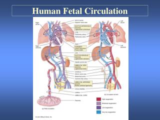
Fetal Circulation. Mike Clark, M.D. Ductus arteriosus. Aorta. Arterial end. Arterial end. Superior vena cava. 4a. Pulmonary trunk. 4. Tubular heart. Ventricle. Foramen ovale. 3. Atrium. 2. Ventricle. 1. Ventricle. Inferior vena cava. Venous end. Venous end. (a) Day 20:

E N D
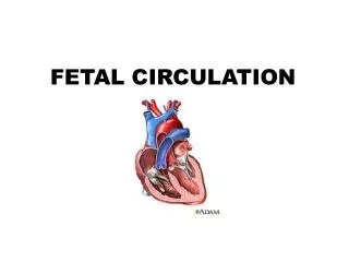
Fetal Circulation Mike Clark, M.D.
Ductus arteriosus Aorta Arterial end Arterial end Superior vena cava 4a Pulmonary trunk 4 Tubular heart Ventricle Foramen ovale 3 Atrium 2 Ventricle 1 Ventricle Inferior vena cava Venous end Venous end (a) Day 20: Endothelial tubes begin to fuse. (b) Day 22: Heart starts pumping. (c) Day 24: Heart continues to elongate and starts to bend. (d) Day 28: Bending continues as ventricle moves caudally and atrium moves cranially. (e) Day 35: Bending is complete. Figure 18.23
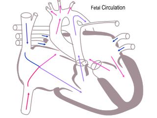
1. Oxygenated blood comes from the placenta traveling to the fetus in the umbilical Vein 2. The umbilical vein dumps 50% of blood into the inferior Vena Cava – through the Ductus Venosus and the other 50% Of the blood enters the inferior Margin of the liver 3. The blood from the inferior Vena Cava enters into the right Atrium 4. Since the placenta is supplying Oxygen to the fetus – much blood is Shunted away from the lungs through The Foramen Ovale and the Ductus Arteriosus. Blood travels from the left ventricle to the Aorta – which gives blood to perfuse the organs. 6. Blood then enters the very low resistance umbilical arteries on its way back to the placenta. Figure 28.13a
In the fetal period- pulmonary vascular resistance is high causing right sided heart pressure to be higher than left sided heart pressure. Thus blood Is shunted from the right side to the left through the Foramen Ovale and Ductus Arteriosus. The Foramen Ovale is an opening between the right atrium and left. The Ductus Arteriosus is an opening between the pulmonary artery and aorta.
Respiratory Transition at Birth • With birth the following sequence occurs: • The placental circulation is removed and systemic vascular resistance increases. This in turn, increases pressures in the left ventricle and left atrium. • The foramen will tend to close when pulmonary blood flow increases as increased pulmonary flow will increase the blood volume entering the left atrium and in so doing, increase left atrial pressure • Increased left atrial pressure (more than right atrial pressure) results in functional closure of the foramen ovale • With the onset of ventilation, the oxygen tension in the alveolus and in arterial blood increases. • as alveolar PaO2 increases, pulmonary vasoconstriction relaxes and pulmonary vascular resistance becomes less than systemic vascular resistance • increased oxygenation also results in constriction of the ductus arteriosus • The onset of ventilation also represents the onset of lung expansion resulting in the straightening out of mechanically compressed vessels • The end result of these changes is a closure of the fetal conduits that carried blood by the lungs, but not into them
The Ductus Arteriosus after closure becomes the Ligamentum Arteriosum The Foramen Ovale after closure becomes the Fossa Ovalis Closure of the Ductus Venosus becomes the Ligamentum Venosum Figure 28.13b
Closure of the Foramen Ovalis • Normally this opening closes in the first three months following birth. When the lungs become functional at birth, the pulmonary pressure decreases and the left atrial pressure exceeds that of the right. This forces the septum primum against the septum secundum, functionally closing the foramen ovale forming the fossa ovalis. In time the septa eventually fuse, leaving a remnant of the foramen ovale, the fossa ovalis. Clinical relevance • In about 30% of adults the foramen ovale does not close completely, but remains as a small patent foramen ovale which gives an atrial septal defect.
Closure of the Ductus Arteriosus The lungs release bradykinin(vasodilator) and falling prostaglandinlevels cause constriction of the smooth muscle in wall of the DA, reducing flow. Usually, the DA begins to close when breathing is established, and is completely sealed after four to ten days. A cord-like vestige of the DA, called the ligamentum arteriosum, remains to connect the exterior of the left pulmonary arteryto the exterior of the aortic arch.
A patent ductus arteriosus allows a portion of the oxygenated blood from the left heart to flow back to the lungs by flowing from the aorta (which has higher pressure) to the pulmonary artery. If this shunt is substantial, the neonate becomes short of breath: the additional fluid returning to the lungs increases lung pressure to the point that the neonate has greater difficulty inflating the lungs. This uses more calories than normal and often interferes with feeding in infancy. This condition, as a constellation of findings, is called congestive heart failure
If Foramen Ovalis does not close then have an Atrial Septal Defect with shunting of blood from left atrium to right atrium due to increased pressure of left atrial pressure over the right atrial pressure.
Sometimes there is a need to temporarily reopen the fetal circulation like In hypoplastic lungs.
Hypoplastic Lungs Pulmonary hypoplasia is part of the spectrum of malformations characterized by incomplete development of lung tissue. Chest radiograph of a newborn with primary pulmonary hypoplasia of the right lung showing shift of the mediastinum to the right hemithorax. Chest radiograph of a newborn with primary pulmonary hypoplasia of the right lung showing shift of the mediastinum to the right hemithorax.