Download
1 / 38
380 likes | 383 Views
This lecture provides an overview of the microscopic structure and development of the central and peripheral nervous system. Topics include the structure of gray matters in the CNS, meninges, ganglia, and peripheral nerves. The text language is in English.

E N D
Lecture 9 General Medicine_3rd semester • MICROSCOPIC STRUCTURE AND DEVELOPMENT OF THE CENTRAL AND PERIPHERAL NERVOUS SYSTEM • Structure of gray matters in the CNS: Iso- and allocortex,cerebellar cortex, spinal cord • Meninges • Ganglia and peripheral nerves • Overwiev of development of the brain and spinal cord including histogenesis of the neural tube
two divisions of the nervous system are distinguished: • - the central nervous system (CNS) - includes the spinal cord and brain • - the peripheral nervous system (PNS) - involves peripheral nerves and ganglia=small aggregations of neurons associated with cerebrospinal or autonomic nerves • Histologically, the nervous system consists of three structurally different • components: • the nerve tissue • blood vessels: capillaries, arterioles and venues that densely penetrate the nerve tissue, • the connective tissuethat serves to protection; it may be differentiated into: • - meninges - forming envelopes of the CNS • - epi-, peri- and endoneurium - connective tissue occurring within nerves or on their surfaces • - capsules surrounding the ganglia
Basic structural characteristics of the nerve tissue • it is composed of • the nerve cells or neurons and special supporting cells called neuroglia • neurons - cells in which two properties of protoplasm are developed to a great degree: irritability (the capacity for response to physical and chemical agents with the initiation of an impulse), conductivity (the ability to transmit impulses from one locality to another) • the cell body or perikaryon • dendrites (their number varies in a great range, theoretically from one to several hundreds; they are usually short and conduct impulses to the perikaryon) • the axon (neurite) - it is mostly very long and always single, it conducts the impulses away from the respective cell. • twig-like branchings or arborizations (telodendria) that touch the perikarya, dendrites or axons of one or more neurons synapses
Synapses unidirectional transmission of signals synapses chemical electric Chemical synapse • presynaptic terminal (knob) • synaptic cleft • postsynaptic terminal (membrane)
neuroglial cells are classified - central glia: astrocytes, oligodendrocytes, microglia and ependyma - peripheral glia: cells of Schwann, satellitecells Astrocytes - are the largest of neuroglial cells, are the protoplasmic and the fibrous ones, both cell types send off numerous processes that extend to blood vessels and to neurons where they expand (end feet) hematoencephalic barrier the protoplasmic astrocytes prevail in gray matter while the fibrous ones in white matter
Oligodendrocytes- are found in particular in white matter where are arranged in rows between the myelinated fibres oligodendrocytes produce the myelin of myelinated axons in white matter Microglia - are the smallest of all glial cells they have smaller and elongated cell bodies that are continuous with one or two thick processes that branch freely in gray matter is of mesenchymal origin and is phagocytic
Ependyma - forms a lining of ventricles and the spinal canal it consists of cubical or columnar cells the eponym is closely associated with pia mater that is extremely vascular and forms so-called choroids plexus Schwann's cells - accompany myelinated fibres in peripheral nerves Satellite cells - are found in spinal or autonomic ganglia where surround cell bodies of neurons in a single layer
THE CENTRAL NERVOUS SYSTEM CNS involves the spinal cord and brain brain: brain stem (myelencephalon, metencephalon, mesence- phalon, diencephalon) cerebellum telencephalon are consisted of two matters differing in the appearance and structure thegray and thewhite matter
the gray matter contains cell bodies, nonmyelinated fibres, associated neuroglial cells (astrocytes, microglia) and very dense capillary network the white matter is composed of only myelinated axons of nerve cells plus oligoden-drocytes and blood capillaries (lesser than in gray matter) Distribution of the gray and white matter spinal cord - the gray matter occurs in the core of the organ, resembling the general form of an H on cross sections (the white matter is peripherally) cerebrum and cerebellum- the gray matter is superficially forming an outer cover designated as the cortex, the white one occupies the inner core islands of the gray matter scattered the white matter in the centre are nuclei
Description of gray matters in the CNS 4 locations: as the gray matter of the spinal cord, as the cerebellar cortex, as the cerebral cortex (telencephalon), and as the cerebellar and cerebral nuclei SPINAL CORD the gray matter is placed centrally and resembles the H-shaped area on each side, the limbs of the capital H are termed the anterior and posterior horns in addition, extending throughout the thoracic segments and first lumbar ones, there are lateral horns of gray matter central canal, lined by ependyma, is situated in the horizontal bar of the H
the gray matter the white matter 3 fasciculi (anterior, lateral and posterior)
Nerve cells of the gray matter are multipolar and of 3 types: • motor neurons- in the anterior (ventral) horns; stellate shape, 150 um in diameter and send off long and myelinated axons ending on muscle fibres of striated voluntary muscles • funicular cells (cellulae funiculares) - mainly in the posterior horns; the axons of the funicular cells enter the white matter and run to the brain stem where end • interneurons (intercalated neurons) - small neurons diffusely distributed among motor and funicular cells
CEREBELLUM vermis and 2 hemispheres surface area is cca 0,10 - 0,15 m2 gray matter occurs in 2locations: a thin cortex at the surface of cerebellar hemispheres and within the centrally placed white matter where forms several small collections of nerve cells called the cerebellar nuclei white matter: fills spaces between cortex and nuclei on sections, it forms „arbor vitae“
in sections, the cerebellar cortex shows 3-layered structure: • an outer molecular layer(stratum moleculare) • Purkinje cell layer(stratumgangliosum) • granule cell layer(stratum granulosum)
efferent fibers - axons of • Purkinje cells • afferent fibers of 2 types: • mossy fibres(they form synapses on cells of granule cell layer) • climbing fibers (take part in synapses on dendrites of Purkinje cells)
TELENCEPHALON gray matter: • cortex telencephali (on the surface) • nuclei thicknes of the cortex 1,5 - 5 mm, area 0,20 – 0,25 m2 white matter: between cortex and nuclei Cortex telencephali/cerebri) • isocortex- asi 11/12 • allocortex Isokortex (neocortex) 8–9 milliard cells Neurons: - pyramidal cells - fusiform cells - stellate (granule) cells - special neurons:horizontal cells (cells of Cajal) and vertical cells (cells of Martinotti)
The axons of cells in the outer pyramidal layer, ganglionic layer and polymorphic (fusiform)layer leave the cortex, enter the white matter and connect different regions of the cortex in the same hemisphere - association fibres or connect areas of the cortex of one hemisphere with corresponding areas of opposite hemisphere - commissural fibres or connect the cortex with lower nervous centres - projection fibres Maps of the izocortex Although 6-layered organization is found in the whole isocortex, the thickness of individual layers and their relative proportions as well as density (total number) of neurons, glial cells, fibers and blood vessels in them show more or lesser conspicuous differences among cerebral lobes and gyruses the differences have been extensively studied in 30 and 40 years of last century on serial sections of cerebral hemispheres and resulted in construction of special maps of the isocortex Maps are termed according to information involved in them as follows: • cytoarchitectural maps - describe the density of perikarya, • myeloarchitectural maps - show the density of myelinated fibers, • glioarchitectural maps - concern the type and density of glial cells, • angioarchitectural maps - describe the density of blood capillaries or vascu-larization, • synaptoarchitectural maps - show the density synapses in the isocortex Allocortex showsmore simple structure than the isocortex, it approximately corresponds to primitive part of telencephalon known as the rhinencephalon
MENINGES • The CNS is protected from the external trauma by bony encasements (skull and vertebral column) and by three membranous investments. • the outermost, robust dura mater • the middle, spider web-like arachnoid and • the innermost, delicate, vascular pia mater • Dura mater • includes: a) the cranial dura, b) the spinal dura • The cranial dura consists of two layers: • - an outer endosteal layer of dense connective tissue, adhering to the inner surface of the bones of the skull • - an inner meningeal layer, consisting of a thinner fibrous tissue membrane, which is covered on its inner surface by mesothelial cells. • These two layers separate each other at certain locations to form the extensive venous (dural) sinuses. • The spinal dura is a continuation of the inner layer of the cranial dura.
Arachnoid is a delicate, avascular membrane lying immediately beneath the dura it consists of 2 components: - a thin, connective tissue component being in contact with the dura - a network of delicate trabeculae, which insert to the pia mater they are covered with flat or low cuboidal epithelium the cavity between the arachnoid and pia mater is a subarachnoid space and is filled with cerebrospinal fluid (CSF) Pia mater the innermost layeris highly vascularized, adheres closely to the brain and spinal cord and follows all of their surface irregularities - inner layer of elastic and reticular fibers, which are firmly attached to the under-lying nervous tissue, - superficial layer receiving the trabeculae from the arachnoid iIts external aspect is covered with simple squamous cells of mesodermal origin Cerebrospinal fluid (CSF) - is a filtrate of blood; it is produced in choroid plexus (folds of pia mater and blood vessels covered with the ependymal cells), located in the ventricles of the brain
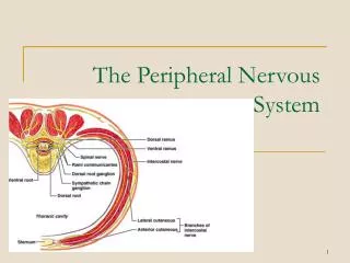
THE PERIPHERAL NERVOUS SYSTEM includes nerves and ganglia of the peripheral nerves NERVES are composed of parallel running nerve fibres and intervening connective tissues are grouped in thin primary nerve bundles with a small amount of loose connective tissue - endoneurium several primary bundles unite to form a secondary nerve bundle surrounded by perineurium in accordance with thickness of the nerve, a few to several tenth of secondary nerve bundles are integrated in the proper nerve enveloped by epineurium that consists of dense connective tissue
GANGLIA ganglia are usually ovoid structures encapsulated similar as nerves by a dense connective tissue two types of ganglia: - dorsal root ganglia (sensory)- are interposed in the course of the dorsal (posterior) roots of the spinal nerves and in some cranial nerves - autonomic ganglia- are associated with nerves of the autonomic nervous system dorsal root ganglia contain the pseudounipolar neurons autonomic ganglia medium-sized multipolar neurons perikarya of neurons are enveloped by a layer of small flat or cuboidal glial cell known assatellite cells
Overwiev of development of the brain and spinal cord including histogenesis of the neural tube it develops from a thickened area of the embryonic ectoderm - neural plate it occurs very early on the dorsal aspect of the embryonic disc cranially to the primitive knob reaching to the oropharyngeal membrane over the notochord
on about day 18, the neural plate begins to invaginate along the cranio-caudal axis and forms neural groove limited with neural folds on each side by the end of the third week, the neural folds become to move together and fuseinto a neural tube the neural tube separates from the ectoderm and is then located between it and notochord neural folds at the time when the neural folds fuse, some neuroectodermal cells separate from them and form along the dorsal aspect of the tube single cord - called the neural crest; it soon divides in the left and right parts that migrate to the dorsolateral aspect of the neural tube neural crest cells give rise to cells of the spinal ganglia and cells of the autonomic ganglia neural groove spinal ganglion neural tube
from the beginning, the proximal segment of the neural tube is broadened and corresponds to future brain the narrower caudal one develops in the spinal cord
Histogenesis of the neural tube the wall of the neural tube is initially composed of a thick, pseudo-stratified columnar epithelium cells then rapidly proliferate in entire thickness of the wall, but later the mitotic activity is reduced only on cells situated near the luminal aspect of the neural tube; as a result of this process, the wall of neural tube differentiates into 2 zones: the inner germinative and the outer marginal ones in the germinative zone the cells continue in their mitotic activity and migrate peripherally. finally, the wall of neural tube shows 3-layered structure: - the ependymal layer = ependyma, - the intermediate or mantle layer= gray matter -cells of mantle layer soon differentiate into primitive neurons - neuroblasts and spongioblasts (glioblasts), -the marginal layer = white matter (contains no cells)
DEVELOPMENT OF THE SPINAL CORD it develops from the caudal portion of the neural tube in contrast with lateral walls of the neural tube, where cells rapidly proliferate, the dorsal and ventral aspects remain thin longitudinal groove - sulcus limitans - divides both lateral walls in the dorsal part - alar plate and ventral part - basal plate cells of mantle layer rapidly proliferate and differentiate in the gray matterRemember: The alar plate - gives rise to dorsal horn, the basal plate - to ventral horn
Positional changes of the spinal cord Initially, the spinal cord extends the entire length of the vertebrate canal during further development, the vertebrate canal grows rapidly than spinal cord and its caudal end gradually comes to lie at relatively higher levels in adults, it usually terminates at the inferior border of the first lumbar vertebra DEVELOPMENT OF THE BRAIN the brain develops from the cranial part of the neural tube at the fourth week,three primary brain vesicles occur: - the forebrain - prosencephalon - the midbrain - mesencephalon - the hindbrain - rhombencephalon During the fifth week, the forebrain and hindbrain divides so that 5 secondary vesicles arise: seediagram (it includes information concerning the development of cavities)
TELENCEPHALONVENTRICULI LAT.CEREBRI PROSENCEPHALON DIENCEPHALON VENTRICULUS TERTIUS MESENCEPHALON MESENCEPHALON AQUAEDUCTUS CEREBRI METENCEPHALON RHOMBENCEPHALON VENTRICULUS QUARTUS MYELENCEPHALON