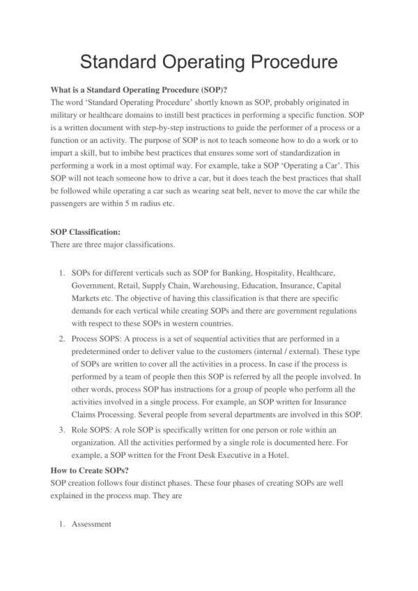
Standard Operating Procedure
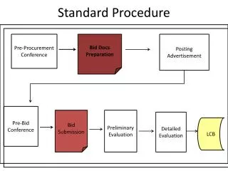
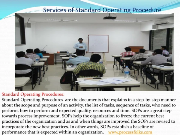
The word ‘Standard Operating Procedure’ shortly known as SOP, probably originated in military or healthcare domains to instill best practices in performing a specific function. SOP is a written document with step-by-step instructions to guide the performer of a process or a function or an activity. The purpose of SOP is not to teach someone how to do a work or to impart a skill, but to imbibe best practices that ensures some sort of standardization in performing a work in a most optimal way. For example, take a SOP ‘Operating a Car’. This SOP will not teach someone how to drive a car, but it does teach the best practices that shall be followed while operating a car such as wearing seat belt, never to move the car while the passengers are within 5 m radius etc. SOP Classification: There are three major classifications. 1. SOPs for different verticals such as SOP for Banking, Hospitality, Healthcare, Government, Retail, Supply Chain, Warehousing, Education, Insurance, Capital Markets etc. The objective of having this classification is that there are specific demands for each vertical while creating SOPs and there are government regulations with respect to these SOPs in western countries. 2. Process SOPS: A process is a set of sequential activities that are performed in a predetermined order to deliver value to the customers (internal / external). These type of SOPs are written to cover all the activities in a process. In case if the process is performed by a team of people then this SOP is referred by all the people involved. In other words, process SOP has instructions for a group of people who perform all the activities involved in a single process. For example, an SOP written for Insurance Claims Processing. Several people from several departments are involved in this SOP. 3. Role SOPS: A role SOP is specifically written for one person or role within an organization. All the activities performed by a single role is documented here. For example, a SOP written for the Front Desk Executive in a Hotel. How to Create SOPs? SOP creation follows four distinct phases. These four phases of creating SOPs are well explained in the process map. They are 1. Assessment 2. Development 3. Implementation 4. Continuous Improvement Assessment: The activities involved during this phase are identifying a sponsor, creating a project charter, management buy-in, identifying the primary, secondary and management processes, identifying the stakeholders and defining the need for SOP. This is like a planning and preparatory phase in SOP creation. Development: During this phase the process maps for the various processes identified during the assessment phase is developed and the SOPs are chiselled. This is a very intense phase, where lots of elicitation activities happen within the organization (Elicitation is the process of drawing out the requirements from the stakeholders). Implementation: The developed SOPs are reviewed during this phase and several training sessions are conducted to move the organization from the current state to the future state. This is also a change management stage where the practices are corrected to attain a state of best-practices. Continuous Improvement: SOP is not a static document, but rather a living document. It has to keep abreast of the various development that undergoes in the industry. The situations when a SOP need to undergo change are typically chance in customer preference or behaviour, changes in regulatory framework, government policies, market dynamics or disruptions etc. The department or role responsible for the SOP need to sense these need for changes and appropriately take action and update the SOP. An SOP that is considered as done and no improvement is carried out has a very short shelf life. Benefits of Implementing SOP SOPs are the documents that help organizations to make the best practices as a part of organizational culture in performing the various processes, functions and activities. The macro-level benefits of SOP are • Well defined steps to perform a work • Standardization of activities irrespective of who is performing • Improved safety and security in operation • Best quality is delivered consistently in a repeated manner • Easy to train new joinees • Sets a standard on expected performance • Minimizing wastages in processes • A platform for continuous improvement • A document for management intentions in Court of Law. About the Author: Mr. Venkadesh Narayanan is the Principal Consultant at Fhyzics Business Consultants Private Limited. He is a Mechanical Engineer and an MBA with over 20 years of experience in Consulting, Business Analysis and Process Improvement. Mr. Narayanan is a former member of Indian Civil Services [IRAS 2000 Batch] and served at Indian Railways, Larsen & Toubro–ECC, Siemens (USA), Euro-Pro LLC (USA) and Latex International (USA) prior to joining Fhyzics. He is also a member of several professional bodies and holds the below certifications: • Certified Business Analysis Professional™, IIBA®, Canada • Certified PMI - Professional in Business Analysis (PMI-PBA)®, USA • Certified Packaging Professional (CPP), IoPP, USA; • Certified Business Process Professional (CBPP®), ABPMP, USA • Certified in Production and Inventory Management (BSCM), APICS, USA; • Certified in Lean from Society of Manufacturing Engineers, USA; • Certified in Six Sigma from Motorola University, USA; If you know more information Please visit: www.processfolks.com
641 views • 4 slides