Download
1 / 6
100 likes | 435 Views
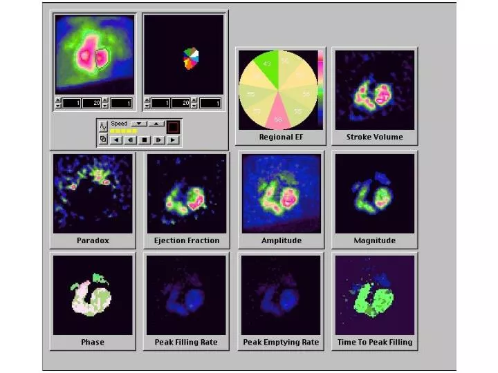
Multiple gated ventriculography, MUGA. Imaging the blood-pool of the heart by synchronizing scintigraphic recording with electrocardigram Repetitively sampling of a specific phase of the cardiac cycle from each of many cycles until an image of reasonable count density is recorded

E N D
Multiple gated ventriculography, MUGA • Imaging the blood-pool of the heart by synchronizing scintigraphic recording with electrocardigram • Repetitively sampling of a specific phase of the cardiac cycle from each of many cycles until an image of reasonable count density is recorded • Evaluation of both global and regional ventricular function
Multiple gated ventriculography, MUGA • Radiopharmaceuticals • Tc-99m RBC labeling • Tc-99m human serum albumin (HSA)
Infarct-avid scintigraphy • Tc-99m Pyrophosphate, PYP • 臨床應用 • 急性心肌梗塞發病後10~12小時內,病灶即可顯示,對病情估計和預後極有幫助。 • 穿壁性病灶在發病2週內的陽性率為90%左右,二週後逐漸轉為陰性。 • 本法對心電圖和 學檢查結果分析有困難的患者診斷價值尤為突出。 • 但心內膜下心肌梗塞的陽性率較低,約60%左右。 • 心肌彌漫性放射性集聚多不是急性心肌梗塞。 • 注意識別骨骼和胸壁、乳房、心包膜、心瓣膜的鈣化灶影像,可大大減低假陽性。 • 本法能夠鑑別急性和陳舊性心肌梗塞,對發現在陳舊性心肌梗塞基礎上發生的再梗塞極有價值。
Cardiac PET N-13-NH3 PET:休息狀態下左心室側壁呈現灌流缺損。 F-18-FDG PET:同部位可見心肌葡萄糖代謝增強。代表此為慢性缺血區域,心肌細胞仍然存活。 此類病人需積極給予血管再通術,方能提高病人存活率與生活品質。