Download
1 / 45
460 likes | 884 Views
Idiopathic Facial Palsy. Bi guo rong. Definition. Idiopathic Facial palsy is also called Bell`s palsy or Facial neuritis. It is characterized by a rapid onset of facial palsy of peripheral type. Etiology. the cause is unclear yet It may be (1)Chill

E N D
Idiopathic Facial Palsy Bi guo rong
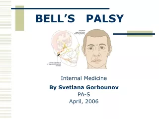
Definition Idiopathic Facial palsy is also called Bell`s palsy or Facial neuritis. It is characterized by a rapid onset of facial palsy of peripheral type
Etiology • the cause is unclear yet • It may be (1)Chill (2)inflammation of virus vasoconstriction----ischemia---edema (3)instability of autonomic nerve
pathology: • swelling of facial nerve • Demyelination and degeneration of the axon
Clinical Features • It may occurs at any age usually in young adults males are commoner then females. The onset is acute. • At the onset there may be pain in the mastoid region or around the angel of the jaw. • The symptoms usually produce its maximum effect within several hours or one to two days. The features is paralysis of the unilateral muscles of expression.
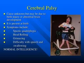
Clinical Features the upper facial paralysis • the wrinkles of the brow are smoothed out Frowning and raising the eyebrow are impossible • the palpebral fissure is wider and closure of the eye is impossible because of the paralysis of the orbicularis
Bell’s phenomenon • the palpebral fissure is wider and closure of the eye is impossible because of the paralysis of the orbicular ,when the patient attempt to close the eye ,the eye globe rolls upwards and slightly out .
Clinical Features the lower facial paralysis • the nasolabial furrow is smoothed out and the mouth is dropped and drawn over to the sound side • the patient is unable to purse the lips and whistle • Owing to paralysis of the buccinator the food tends to accumulate between the teeth and cheek. Dribbling may occur.
Diagnosis and Differential Diagnosis • According to the rapid onset of facial palsy of peripheral type • Differential Diagnosis 1 Guillain - Barre - Syndrome 2 Otitis media. labyrinthitis 3 posterior fossa disease
Treatment Acute phase • prednisone 10mg tid po. 5—10days • VitamineB1.B12 • physiatrists • the protection of the expose cornea
Treatment recovering phase • exercise of the paralyzed facial muscles • acupuncture • physiatrists • facio-hypoglossal anastomosis.
prognosis • The prognosis is usually good. • After one to two week .the patient begin to recover . • After one to two month .the patient recover prominently. • About seventy fine percent of causes can recover completely. • If the patient can not recover in six month, he will suffer from the disability.
Acute inflammatory demyelinating polyneuropathy (AIDP) Guillain---Barre Syndrome
Definition It is a rapidly progressive polyneuropathy. The damaged region is usually the spinal roots and peripheral nerves, occasionally the cranial nerves .the pathologic change is segmental demyelination . The manifestation is characterized by a symmetric tetraplegia.
Etiology • post—infective allergic disease
Pathology • the damaged region is usually the spinal roots and peripheral nerves ,particularly ventral roots, occasionally the cranial nerve. • Segmental demyelination of the spinal roots and peripheral nerves. perivascular inflammatory infiltration mainly is lymphocyte and monocyte .
Clinical Features • The onset is acute or subacute. Before one to four weeks of the onset ,there may be symptoms of the upper airway infection or gastrointestinal infection .
Clinical Features symmetric tetraplegia • it is often the first symptom. The paralysis is flaccid. The muscular tonus is low , tendon reflexes are diminished or lost and pathologic reflexes are negative. When respiratory muscles are affected, respiratory failure may occur.
Clinical Features sensory disturbance • paresthesias : numbness ,tingling ,tenderness . • peripheral sensory impairment: stocking—glove pattern • the sensory symptoms are always present it is slighter than paralysis of the limbs.
Clinical Features cranial nerves paralysis • the paralysis of the facial nerves on both sides • bulbar paralysis • others: trigeminal hypoglossal oculomotor trochlear and abducens nerves .
Clinical Features the symptoms of vegetative nervous systems • sweating , flushing ,swelling and nutritional disturbance of the skin • tachycardia , variety of the blood pressure • the sphincter disturbance is rarely occasionally there may be slight retention of urine.
Clinical Features • The disease usually produces its maximum in one to two weeks. • It is a self—limited—disease. About 85% of patients would have a complete recovery . • the most important complications are : a. respiratory muscles paralysis. b .pneumonia. C .heart failure . when the patients have all these complications, the prognosis is not very good.
Laboratory Aids CSF: • dissociation albumino—cytologique • the change is a great excess of protein with a normal cell count the high protein is most prominent after the third week of the onset and may persist for many weeks after recovery.
Laboratory Aids peripheral blood phase : • the white cells count may be high .
Diagnosis: • The history of a recent infection • The onset is acute or subacute • Symmetric flaccial tetreplegia • Damage of the cranial nerves • CSF: dissociation albumino—cytologique
Differential Diagnosis • Acute Poliomyelitis • Acute Myelitis • Periodic Paralysis • Myasthenic Gravis
Treatment Etiological treatment : • Plasma exchange: 40ml/kg • Intravenous immunoglobulin, IVIG : 0.4g/(kg·d) ,5 days • Corticosteroids : Symptomatic treatment :
treatment • Recovery stage 1.exercise of the limbs 2.acupuncture 3.physiatrics 4.massotheraohy
Definition • Acute myelitis is an acutenon-specificity inflammation of several segments of the spinal cord ,the cause is unkown yet, the manifestation is characterized by that of atransverse lesion of the spinal cord –including motor,sensory and autonomic disturbance.
Etiology • The cause is unkown yet. • Maybe the inflammation of virus or immunization induce the auto-immune reaction.
pathology • To the naked eye :the lesion is edema hyperemia and softening. • Microscopically: the lesion is infiltrated with inflammatory cells ,degenaration of the axon and demyelination.
Clinical features • Acute myelitis often occurs in youth and adults ,the onset is acute ,before a few weeks or after days of the onset ,the infective symptoms of the upper airway may occur ,the earliest symptom is usually the weakness or numbness of the legs.
Clinical features • Acute stage • (1)motor disturbance • Spinal cord shock : • Flaccid paraplegia: the muscular tonus is low , tendon reflexes are diminished or lost ,pathological reflex is negative , retention of urine.
Clinical features • Acute stage • (2)sensory disturbance • All sensations below the level of the lesion are diminished or lost • There may be a zone of hyperaesthesia between the area of the sensory loss and that of normal sensibility
Clinical features • Acute stage • (3)autonomic disturbance • Impairment of sphincter control : the retention of urine; • others : hypohidrosis, dermal edema , dryness
Clinical features • Ascending myelitis : • It is severe when the lesion involves the upper cervical cord and medulla ,tetraplegia and dyspnea may occur ,the patient often dies from respiratory failure ,the mortality is very high.
Clinical features • Recovering stage • After three or four weeks of the onset , the acute stage is over and the patient becomes recovering . The clinic symptoms depends on the severity of the lesion and absence or presence of the complication.
Clinical features • Recovering stage • (1)motor disturbance : the upper motor neuron paraplegia , the muscular power progress gradually ; tendon reflexes are increased; pathological reflexes are present. • (2)sensory disturbance :the level descends gradually ,the recovery of sensory disturbance is slower than that of motor disturbance ; • (3)autonomic disturbance :reflex urination
Laboratory aids • CSF :the CSF is commonly abnormal. it is crystal clear ,the pressure is normal , with lymphocytosis or a slightly increased protein concentration. (especially if examined soon after an acute relapse ) glucose and chloride of CSF are normal . • Peripheral blood phase : the count of WBC is normal or slightly high.
Diagnosis • The rapid onset ; • Premonitory infective symptom ; • The symptoms of a transverse lesion of the spinal cord ; • CSF is slightly abnormal .
Differential diagnosis • Acute inflammatory demyelinating polyneuropathies (AIDP) • Neuromyelitis optiea • Hematomyelia • Acute spinal epidural abscess • Spinal cord compression • TB • Tumor
treatment • DRUG • (1)Corticosteroids: • methylprednisolone 500~1000mg qd iv ,3~5days • dexamethasone 5~10mg qd iv 10days • Prednisone 40~60 mg qd po 1~2months • (2)Intravenous immunoglobulin, IVIG • (3) antibiotics • (4)vitamin B
treatment • Good nursing: • Rehabilitation :physical therapy