Download
1 / 39
430 likes | 651 Views
Electron Microscopes. Gateways to the hidden world of the super small. An electron microscope (EM) is a type of microscope that uses an electron beam to illuminate a specimen and produce a magnified image.

E N D
Electron Microscopes Gateways to the hidden world of the super small.
An electron microscope (EM) is a type of microscope that uses an electron beam to illuminate a specimen and produce a magnified image.
The electron microscope uses electrostatic and electromagnetic lenses to control the electron beam and focus it to form an image. These electron optical lenses are analogous to the glass lenses of a light optical microscope.
The original form of electron microscope, the transmission electron microscope (TEM) uses a high voltage electron beam to create an image.
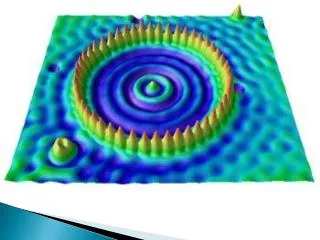
An EM has greater resolving power than a light microscope and can reveal the structure of smaller objects because electrons have wavelengths about 100,000 times shorter than visible light photons. They can achieve magnifications of up to about 10,000,000x
Transmission electron microscopy (TEM) is a technique where a beam of electrons is transmitted through an ultra-thin specimen. An image is formed from the interaction of the electrons transmitted through the specimen. The image is magnified and focused onto a fluorescent screen, or a layer of photographic film, or to be detected by a sensor such as a CCD camera.
The major disadvantage of the transmission electron microscope is the need for extremely thin sections of the specimens, typically about 100 nanometers. All images are 2D. Seen below is a plant cell in the tip of a root.
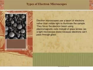
Scanning electron microscope. This scope uses a focused beam of high energy electrons to view the surface of solid specimens. Magnification ranges from 20X to 30,000X.
Surface of an erasable, programmable, read-only memory, silicon microchip
Household dust: includes long hairs of cat fur, twisted synthetic and woollenfibers, serrated insect scales, a pollen grain, and plant and insect remains