Download
1 / 44
890 likes | 2.28k Views
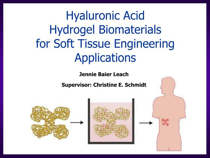
Hyaluronic Acid Hydrogel Biomaterials for Soft Tissue Engineering Applications. Jennie Baier Leach Supervisor: Christine E. Schmidt. “Biomaterials that heal” Ratner BD . (2002) J Controlled Release . 78:211-8. Aim : To facilitate natural wound healing biology. Biological design :

E N D
Hyaluronic Acid Hydrogel Biomaterials for Soft Tissue Engineering Applications Jennie Baier Leach Supervisor: Christine E. Schmidt
“Biomaterials that heal” Ratner BD. (2002) J Controlled Release. 78:211-8 Aim: To facilitate natural wound healing biology Biological design: Biomimetic molecules like those present in a wound Limited nonspecific protein adsorption Nonimmunogenic Materials design: Enzymatic degradation Versatile modification strategies Mechanical properties match tissue www.organogenesis.com Hubbell JA. (1999) Curr Opin Biotech. 10:123-9; Stocum DL. (1998) Wound Repair Regen. 6:276-90
Tissue Engineering Scaffolds: State of the Art Proteins Fibrin, Collagen “Sugars” Agarose, Alginate Chitosan, Dextran, Hyaluronic acid Synthetic polymers Polyethylene glycol (PEG) Polylactic-co-glycolic acid (PLGA) Polyhydroxyethyl methacrylate (pHEMA) Inherent biological activity Nonimmunogenic Multiple modification sites Tunable material properties
Hyaluronic Acid (HA) glucuronic acid acetylglucosamine • Natural ECM component • Easily produced in large quantities • Non-immunogenic • Multiple sites available for modification • Enzymatically degradable
HA’s role in wound healing Chen WY & Abatangelo G. (1999) Wound Repair Regen. 7:79-89.
Overall Goal: To develop and characterize hyaluronic acid hydrogel scaffolds for soft tissue engineering applications
Aim 1: To create and characterize HA hydrogels Aim 2: To develop methods for controlling cell-HA hydrogel interactions Peptide-conjugated HA hydrogels Protein-releasing HA hydrogels
Aim 1: To create and characterize HA hydrogels Aim 2: To develop methods for controlling cell-GMHA hydrogel interactions Peptide-conjugated GMHA hydrogels Protein-releasing GMHA hydrogels
glycidyl methacrylate (GM) + Photoinitiator + UV light Crosslinked GMHA GMHA HA GM modification HA Methacrylation and Crosslinking Baier Leach J, Bivens KA, Patrick CW, Jr. & Schmidt CE. (2003) Biotech Bioeng 82:578-89 Crosslinking Variables: GMHA concentration (0.5-2.0%) UV exposure (1-4 min, ~22 mW/cm2) Photoinitiator conc. (0.03-3% Irgacure 2959) % methacrylation (NMR: ~5-11%)
GMHA Solution +UV Remove mold Equilibrate in buffer overnight, weigh Dry completely, weigh Determining the Degree of Crosslinking (Swelling Ratio) Goal: To obtain a relative measure of crosslinking for the GMHA gels
Flory polymer-solvent interaction theory: Estimated pore size: 644 nm 619 nm 539 nm • % methacrylation • Swelling ratio Cross-linking • Pore size Effect of Methacrylation on Crosslinking n=3 for each bar
5% 7% 11% Increasing % methacrylation n>4 for each point Effect of Methacrylation on Degradation Rate GMHA Solution +UV Incubate in hyase Measure weight loss of gel over time Remove mold
Media Hydrogel Crosslinked GMHA 1% GMHA 0.1% Irgacure 2959 0.03% N-vinyl pyrrolidinone 1 minute UV Crosslinked GMHA GMHA in solution Media HA n>6 for each bar Effect of Methacrylation on Cytocompatibility HAEC monolayer www.corning.com
3. Harvest tissue at 2 weeks (side view without TEC) 1. Fill TEC with hydrogel 4 hydrogels per rat + Control: Fibrin -Control: Agarose 2 HA gels 2. Suture to muscle 4. EC immunostain (CD31) In Vivo Analysis of Endothelial Cell (EC) Infiltration
In Vivo Analysis of EC Infiltration Fibrin (+) GMHA hydrogel 6.63 ± 1.10, n = 9 (% area CD31-positive cells)7.06 ± 0.14, n = 4 2 Week Implant Scale, 200 mm Agarose (-)
Aim 1: To create and characterize HA hydrogels Aim 2: To develop methods for controlling cell-GMHA hydrogel interactions Peptide-conjugated GMHA hydrogels Protein-releasing GMHA hydrogels
Aim 1: To create and characterize HA hydrogels Aim 2: To develop methods for controlling cell-GMHA hydrogel interactions Peptide-conjugated GMHA hydrogels Protein-releasing GMHA hydrogels
Peptide-Conjugated GMHA Hydrogels Baier Leach J, Bivens KA, Collins CN, & Schmidt CE. (submitted) J Biomed Mater Res * Peptides of interest: Cell adhesive fibronectin peptide (GRGDSG) Model peptide, hexaglycine (GGGGGG) * Based on work with PEG hydrogels in J. West and J. Hubbell’s laboratories
Conjugation Efficiency and Yield Hexaglycine input Conjugation efficiency Hexaglycine input Conjugation yield Targeted yield * * Based on work with PEG hydrogels in J. West and J. Hubbell’s laboratories
Negative control (no hyase) Effect of [PEG] on Degradation 5 u/ml hyase n>3 for each point Incubate in hyase Measure weight loss
Human dermal fibroblasts 3 days Rinse Count number of adherent cells n=3 hydrogels for each bar Fibroblast Adhesion on GMHA-PEG-GRGDSG hydrogels
Aim 1: To create and characterize HA hydrogels Aim 2: To develop methods for controlling cell-GMHA hydrogel interactions Peptide-conjugated GMHA hydrogels Protein-releasing GMHA hydrogels
Growth factors: ~5-20 nm BSA: ~30 nm 4-arm PEG Density of polymer or crosslinks Protein diffusion ~50 nm Protein-Releasing GMHA hydrogels Baier Leach J & Schmidt CE. (in preparation) Biomaterials ~500 nm
Fractional release of BSA (Mt/M) n=3 for each point BSA Release From GMHA-Based Hydrogels At each time-point: remove 1 ml, measure protein concentration 15ml
At each time-point: remove 1 ml, measure protein concentration 15ml BSA Release From GMHA-Based Hydrogels Fractional release of BSA (Mt/M) Fractional release of BSA (Mt/M) n=3 for each point
Crosslink/Polymer Density Affects More Than Diffusion ~50 nm Density of polymer or crosslinks Protein diffusion Degradation rate Stiffness
Hydrogel-Microsphere Composites BSA-PLGA microspheres (SEM) Dv,10 = 8 mm Dv,50 = 16 mm Dv,90 = 34 mm Hydrogel-microsphere composite (brightfield) 100 mm
Extended BSA Release from Hydrogel-Microsphere Composites Fractional release of BSA (Mt/M) 2 weeks n=3 for each point
Aim 1: To create and characterize HA hydrogels Aim 2: To develop methods for controlling cell-GMHA hydrogel interactions Peptide-conjugated GMHA hydrogels Protein-releasing GMHA hydrogels
Overall Goal: To develop and characterize hyaluronic acid hydrogel scaffolds for soft tissue engineering applications
Acknowledgements Dr. Charles W. Patrick, Jr, MD Anderson Cancer Center, Houston, TX Dr. C.P. Pathak, Sulzer Biologics, Austin, TX Dr. Brent Iverson, UT-Austin Dr. Nicholas Peppas, UT-Austin Dr. Donald Paul, Timothy Fornes, UT-Austin Drs. Keith Johnston, Robert O. Williams III, W. Thomas Leach, UT-Austin Kathryn Bivens, Scott Zawko, Schmidt group members Eric Brey, Cindy Frye, Carol Johnston, Patrick group members Chelsea Collins, Kate Lee, Gwang-Yi Hwang, Erik Askenasy, Nabilla Porbandarwalla, undergraduate research assistants
Cross-linked HA hyase hyase Crosslinked HA Hydrogels Native HA hyase hyase
Previous Work: HA Composite Materials Collier J, Hudson TW, Schmidt CE. (2000) JBMR 50:574 Polypyrrole without HA Polypyrrole with HA ~2 fold increase in blood vessels with HA
Effect of Methacrylation on HAEC Proliferation Set-up: “starve” 12h; expose to 1.5 mg/ml fragments for 48h n>9 for each bar
Flory Polymer-Solvent Theory Qv determined from Qm: x: where Mc from simplified Flory-Rehner equation: therefore, ue: • Qv volumetric swelling u specific volume of the dry polymer n # of monomer repeats • Qm mass swelling Mc average MW between crosslinks x mesh size • rp density of dry polymer V1 molar volume of solvent (water) • rs density of solvent (water) ue effective crosslink density • Flory polymer-solvent interaction parameter (assumed to be 0.473 for HA) • (ro2)1/2root mean square distance between crosslinks
In vivo degradation Muscle or cellular ingrowth 4 weeks 8 weeks Scale, 100 mm H&E stain GMHA hydrogel Gels degrade slowly due to low subcutaneous concentration of hyase
Efficiency and Yield Hexaglycine / gel reactive sites Conjugation efficiency Hexaglycine / gel reactive sites Conjugation yield
Effect of Peptide Conjugation on Crosslinking • Measure swelling ratio • Swelling ratio Crosslinking n>2 for each point
Release Data Fits Fickian Model of Diffusion for Mt/M∞ < 0.6, n=3 for each point; n=9 for De calculations
Diffusion Coefficient of BSA n=9 for each bar
Protein stability Collect BSA released from hydrogels Run SEC/HPLC to determine relative amounts of monomeric BSA and oligomeric BSA aggregates n=4 gels, sampled 3 times each