Download
1 / 41
410 likes | 717 Views
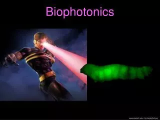
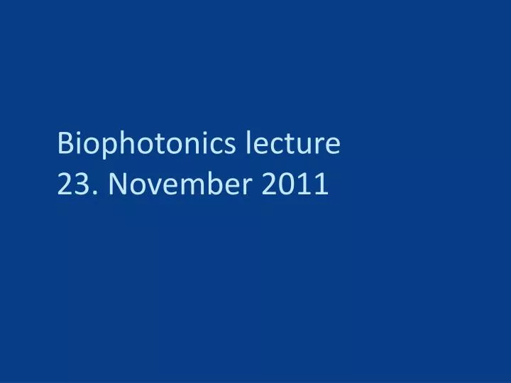
Biophotonics lecture 23. November 2011. Last week: Fluorescence microscopy in general, labeling, etc… How to do optical sectioning and fill the missing cone, the confocal microscope. Today: Structured illumination: an alternative approach to optical sectioning

E N D
Last week: • Fluorescence microscopy in general, labeling, etc… • How to do optical sectioning and fill the missing cone, the confocal microscope
Today: • Structured illumination: an alternative approach to optical sectioning • Super resolution techniques: beyond the Abbe limit • High-resolution structured illumination microscopy
Optical sectioning alternatives: structured illumination microscopy (SIM) • Sample is illuminated with a structured illuminationpattern, i.e. a line grid. • This can be produced by placing a grid in a conjugate image plane in the illumination pathway. • Three images are taken for three different (lateral) positions of the illumination grid. • These images can be computed into a final, optically sectioned image.
z Full-field illumination Structured illumination y y y x x x In-focus sample slice x Out-of-focus sample slice Acquired structured illumination image Acquired wide-field image
Acquired structured illumination images I1(r) I2(r) I3(r) Ifinal(r) = max{I1(r) , I2(r) , I3(r)}- min{I1(r) , I2(r) , I3(r)} Result: sectioned image Wide-field image
Advantages: • Cheaper: no need for laser, scanner, PMTs, AOTFs, etc. • Potentially faster: for a large field of view 3 wide-field images can be acquired faster than a point-wise scan. • Less photo-bleaching: less light is lost as compared to using a pinhole
Frequency support of sectioning SIM Sectioning SIM Wide-field But this isNOT an OTF !! • Filled missing cone • Higher axial support • Higher lateral support (1 direction), more on this later
Wide-field Confocal Sectioning SIM The image formation in section SIM cannot be written as a convolution of the sample with an intensity PSF.
Advantages: • Cheaper: no need for laser, scanner, PMTs, AOTFs, etc. • Potentially faster: for a large field of view 3 wide-field images can be acquired faster than a point-wise scan. • Less photo-bleaching: less light is lost as compared to using a pinhole • Disadvantage: • Not a linear imaging modality. Not as useful for quantitative analysis.
Limited resolution in conventional, wide-fieldimaging Fourier space Real space “Sample“ forsimulation Fourier transform of “Sample“ Sample will be “repainted” with a blurry brush rather than a point-like brush.
high frequencygrid low frequencymoiré patterns Moiréeffect high frequencydetail
Moiréeffect Illumination Sample Structured Illumination Microscopy Illumination withperiodic light pattern down-modulates high-frequency sample informationandmakesitaccessiblefordetection.
x z CCD Tubelens Tubelens Filter Dichromaticreflector Laser Diffractiongrating, SLM, etc… Objective Sample
Structured Illumination Micropscopy Sample withstructuredillumination Illumination Sample Multiplicationof sample andillumination
Structured Illumination Micropscopy Fourier space Real space Convolutionof sample andillumination Multiplicationof sample andillumination
Structured Illumination Micropscopy Illumination Sample
Structured Illumination Micropscopy Sample & llumination Sample
Structured Illumination Micropscopy Sample & llumination Sample Imaging leadstolossof high frequencies (OTF)
Structured Illumination Micropscopy Sample Separatingthecomponents…
Structured Illumination Micropscopy Sample Separatingthecomponents… Shiftingthecomponents…
Structured Illumination Micropscopy Sample Separatingthecomponents… Shiftingthecomponents… Recombiningthecomponents…
Structured Illumination Micropscopy Sample Reconstructed sample Separatingthecomponents… Shiftingthecomponents… • Recombiningthecomponents… usingthecorrectweights.
sample wide-field SIM (x only)
Missingcone – noopticalsectioning 1focus in back focal plane Full-fieldillumination
Missingcone – noopticalsectioning 2foci in back focal plane 2-beam structuredillumination
Missingconefilled – opticalsectioning betterz-resolution 3foci in back focal plane 2-beam structuredillumination
Fourier space (percentile stretch) 1 mm LiisaHirvonen, Kai Wicker, OndrejMandula, Rainer Heintzmann
99 beads averaged wide-field WF: 252 nm SIM: 105 nm SIM
Axon Actin (Growth Cone) 2 µm excite 488nm, detect > 510 nm24 lp/mm = 88% offrequencylimit Plan-Apochromat 100x/1.4 oiliris Samples Prof. Bastmeyer, Universität Karlsruhe (TH)
Axon Actin (Growth Cone) 2 µm excite 488nm, detect > 510 nm24 lp/mm = 88% offrequencylimit Plan-Apochromat 100x/1.4 oiliris Samples Prof. Bastmeyer, Universität Karlsruhe (TH)
Doublets in Myofibrils Isolated myofibrils fromrat skeletal muscle Titin T12 – Oregon green 124 nm 1 µm L. Hirvonen, E. Ehler, K. Wicker, O. Mandula, R. Heintzmann, unpublished results
Molecules in spaceand time living COS1 cell 1 mm L. Hirvonen, K. Wicker., O. Mandulaand R. Heintzmann, Structured illuminationmicroscopyof a livingcell, Europ. Biophys. J. 38, 807-812, 2009
Podosomes 5 mm f-Actin Vinculin Are podosomes arranged as "sticks and joints"? Marie Walde, James Monypenny (cooperation G. Jones), King‘s College London
In Real space In Fourier space Linear Excitation (low intensity) magnitude Support regionof OTF spatial frequency 0 -K0 K0 Non-Linear Excitation (high intensity) magnitude Support regionof OTF -3K0 -K0 0 K0 spatial frequency -2K0
NonlinearStructured Illumination Micropscopy Conventionalmicroscopy Linearstructured illumination Saturatedstructured illumination 1 µm 1 µm 1 µm M.G.L. Gustafsson (2005), PNAS, 37, 13081-13086 Mats Gustafsson, UCSF 50 nm microscpheresnonlinearity: fluorescence saturation, 53J/m2 3 extra harmonics
0.5 µm But: Artefacts possible!
0.5 µm But: Artefacts possible! Sophisticated algorithms are needed!