Download
1 / 1
10 likes | 131 Views
EFFECT OF LOWER-LIMB INJURY ON ULTRASONOGRAPHY MEASURES OF MUSCLE QUALITY AND ARCHITECTURE IN NBA PLAYERS.

E N D
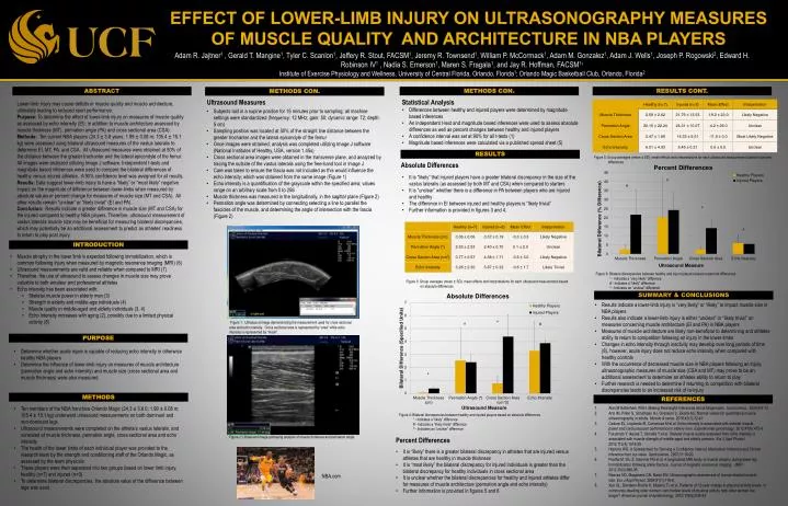
EFFECT OF LOWER-LIMB INJURY ON ULTRASONOGRAPHY MEASURES OF MUSCLE QUALITY AND ARCHITECTURE IN NBA PLAYERS Adam R. Jajtner1 , Gerald T. Mangine1, Tyler C. Scanlon1, Jeffery R. Stout, FACSM1, Jeremy R. Townsend1, William P. McCormack1, Adam M. Gonzalez1, Adam J. Wells1, Joseph P. Rogowski2, Edward H. Robinson IV1 , Nadia S. Emerson1, Maren S. Fragala1, and Jay R. Hoffman, FACSM11 Institute of Exercise Physiology and Wellness, University of Central Florida, Orlando, Florida1; Orlando Magic Basketball Club, Orlando, Florida2 ABSTRACT METHODS CON. RESULTS CONT. METHODS CON. Lower-limb injury may cause deficits in muscle quality and muscle architecture, ultimately leading to reduced sport performance. Purpose: To determine the effect of lower-limb injury on measures of muscle quality as assessed by echo intensity (EI); in addition to muscle architecture assessed by muscle thickness (MT), pennation angle (PA) and cross sectional area (CSA). Methods: Ten current NBA players (24.3 ± 3.8 years; 1.99 ± 0.08 m; 105.4 ± 15.1 kg) were assessed using bilateral ultrasound measures of the vastus lateralis to determine EI, MT, PA, and CSA. All ultrasound measures were obtained at 50% of the distance between the greater trochanter and the lateral epicondyle of the femur. All images were analyzed utilizing Image J software. Independent t-tests and magnitude based inferences were used to compare the bilateral differences of healthy versus injured athletes. A 90% confidence level was assigned for all results. Results: Data suggest lower-limb injury to have a “likely” or “most likely” negative impact on the magnitude of difference between lower-limbs when measured by absolute values or percent change for measures of muscle size (MT and CSA). All other results remain “unclear” or “likely trivial” (EI and PA). Conclusion: Results indicate a greater difference in muscle size (MT and CSA) for the injured compared to healthy NBA players. Therefore, ultrasound measurement of vastus lateralis muscle size may be beneficial for measuring bilateral discrepancies, which may potentially be an additional assessment to predict an athletes’ readiness to return to play post injury. Ultrasound Measures • Statistical Analysis • Differences between healthy and injured players were determined by magnitude based inferences • An independent t-test and magnitude based inferences were used to assess absolute differences as well as percent changes between healthy and injured players • A confidence interval was set at 90% for all t-tests (1) • Magnitude based inferences were calculated via a published spread sheet (5) • Subjects laid in a supine position for 15 minutes prior to sampling; all machine settings were standardized (frequency: 12 MHz; gain: 50; dynamic range: 72; depth: 5 cm) • Sampling position was located at 50% of the straight line distance between the greater trochanter and the lateral epicondyle of the femur • Once images were obtained, analysis was completed utilizing Image J software (National Institutes of Healthy, USA, version 1.45s) • Cross sectional area images were obtained in the transverse plane, and analyzed by tracing the outside of the vastus lateralis using the free-hand tool in Image J • Care was taken to ensure the fascia was not included as this would influence the echo intensity; which was obtained from the same image (Figure 1) • Echo intensity is a quantification of the grayscale within the specified area; values range on an arbitrary scale from 0 to 256 • Muscle thickness was measured in the longitudinally, in the sagittal plane (Figure 2) • Pennation angle was determined by connecting selecting a line to parallel the fascicles of the muscle, and determining the angle of intersection with the fascia (Figure 2) # * RESULTS Figure 5: Group averages (mean ± SD), mean effects and interpretations for each ultrasound measurement based on percent differences Absolute Differences • It is “likely” that injured players have a greater bilateral discrepancy in the size of the vastus lateralis (as assessed by both MT and CSA) when compared to starters • It is “unclear” whether there is a difference in PA between players who are injured and healthy • The difference in EI between injured and healthy players is “likely trivial” • Further information is provided in figures 3and 4. ^ Figure 6: Bilateral discrepancies between healthy and injured players based on percent differences * - Indicates a “very likely” difference # - Indicates a “likely” difference ^ - Indicates an “unclear” difference ^ INTRODUCTION * • Muscle atrophy in the lower limb is expected following immobilization, which is common following injury when measured by magnetic resonance imaging (MRI) (6) • Ultrasound measurements are valid and reliable when compared to MRI (7) • Therefore, the use of ultrasound to assess changes in muscle size may prove valuable to both amateur and professional athletes • Echo intensity has been associated with: • Skeletal muscle power in elderly men (3) • Strength in elderly and middle-age individuals (4) • Muscle quality in middle-aged and elderly individuals (3, 4) • Echo intensity increases with aging (2), possibly due to a limited physical activity (8) Figure 3: Group averages (mean ± SD), mean effects and interpretations for each ultrasound measurement based on absolute differences SUMMARY & CONCLUSIONS Figure 4: Bilateral discrepancies between healthy and injured players based on absolute differences * - Indicates a “likely” difference # - Indicates a “likely trivial” difference ^ - Indicates an “unclear” difference • Results indicate a lower-limb injury is “very likely” or “likely” to impact muscle size in NBA players • Results also indicate a lower-limb injury is either “unclear” or “likely trivial” on measures concerning muscle architecture (EI and PA) in NBA players • Measures of muscle architecture are likely non-beneficial to determining and athletes ability to return to competition following an injury in the lower-limbs • Changes in echo intensity through inactivity may develop over long periods of time (8), however, acute injury does not reduce echo intensity when compared with healthy controls • With the occurrence of decreased muscle size in NBA players following an injury, ultrasonographic measures of muscle size (CSA and MT) may prove to be an additional assessment to determine an athletes ability to return to play • Further research is needed to determine if returning to competition with bilateral discrepancies leads to an increased risk of re-injury Figure 1: Ultrasound image demonstrating the measurement used for cross sectional area and echo intensity. Cross sectional area is represented by “area” while echo intensity is represented by “mean”. PURPOSE • Determine whether acute injure is capable of reducing echo intensity in otherwise healthy NBA players • Determine the influence of lower-limb injury on measures of muscle architecture (pennation angle and echo intensity) and muscle size (cross sectional area and muscle thickness) were also measured. METHODS REFERENCES • Ten members of the NBA franchise Orlando Magic (24.3 ± 3.8 0; 1.99 ± 0.08 m; 105.4 ± 15.1 kg) underwent ultrasound measurements on both dominant and non-dominant legs • Ultrasound measurements were completed on the athlete’s vastus lateralis, and consisted of muscle thickness, pennation angle, cross sectional area and echo intensity • The health of the lower limbs of each individual player was provided to the research team by the strength and conditioning staff of the Orlando Magic, as assessed by the team physician • These players were then separated into two groups based on lower limb injury, healthy (n=7) and injured (n=3) • To determine bilateral discrepancies, the absolute value of the difference between legs was used 1. Alan M Batterham WGH. Making Meaningful Inferences About Magnitudes. Sportscience. 2005;9:6-13. 2. Arts IM, Pillen S, Schelhaas HJ, Overeem S, Zwarts MJ. Normal values for quantitative muscle ultrasonography in adults. Muscle & nerve. 2010;41(1):32-41. 3. Cadore EL, Izquierdo M, Conceicao M et al. Echo intensity is associated with skeletal muscle power and cardiovascular performance in elderly men. Experimental gerontology. 2012;47(6):473-8. 4. Fukumoto Y, Ikezoe T, Yamada Y et al. Skeletal muscle quality assessed from echo intensity is associated with muscle strength of middle-aged and elderly persons. Eur J Appl Physiol. 2012;112(4):1519-25. 5. Hopkins WG. A Spreadsheet for Deriving a Confidence Interval, Mechanisic Inference and Clinical Inference from a p value. Sportscience. 2007;11:16-20. 6. Psatha M, Wu Z, Gammie FM et al. A longitudinal MRI study of muscle atrophy during lower leg Immobilization following ankle fracture. Journal of magnetic resonance imaging : JMRI. 2012;35(3):686-95. 7. Reeves ND, Maganaris CN, Narici MV. Ultrasonographic assessment of human skeletal muscle size. Eur J Appl Physiol. 2004;91(1):116-8. 8. Xue QL, Bandeen-Roche K, Mielenz TJ et al. Patterns of 12-year change in physical activity levels in community-dwelling older women: can modest levels of physical activity help older women live longer? American journal of epidemiology. 2012;176(6):534-43. Figure 2: Ultrasound image portraying analysis of muscle thickness and pennation angle. Percent Differences • It is “likely” there is a greater bilateral discrepancy in athletes that are injured versus athletes that are healthy in muscle thickness • It is “most likely” the bilateral discrepancy for injured individuals is greater than the bilateral discrepancy for healthy individuals in cross sectional area • It is unclear whether the bilateral discrepancies for healthy and injured athletes differ for measures of muscle architecture (pennation angle and echo intensity) • Further information is provided in figures 5 and 6 NBA.com