Download

1 / 29
290 likes | 444 Views
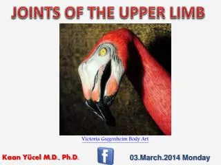
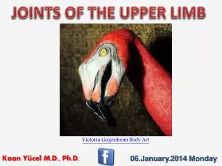
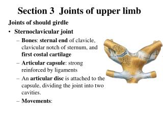
Joints Of Upper Extremities. Sternoclavicular Joint . a saddle joint - the only bony joint between axial skeleton & upper limb clavicle sits in a saddle socket formed by superolateral manubrium & 1st costal cartilage . Sternoclavicular Joint.

E N D
Sternoclavicular Joint • a saddle joint - the only bony joint between axial skeleton & upper limb • clavicle sits in a saddle socket formed by superolateral manubrium & 1st costal cartilage
Sternoclavicular Joint • 1. fibrous capsule - surrounds entire joint, reinforced by ligaments • a. synovial capsule - lines fibrous capsule & both sides of disc • b. lateral & medial synovial membranes line 2 cavities on either side of disc • 2. articular disc - mostly fibrocartilage - strong, thick, very dense • a. attached: superiorly - to medial end of clavicle inferiorly - to junction btw manubrium & 1st costal cartilage • b. continuous with sternoclavicular ligament • c. absorbs shock & prevents medial displacement of clavicle
Sternoclavicular Joint • 3. anterior, posterior sternoclavicular ligaments - thickenings of the fibrous capsule • 4. Interclavicular ligament - also a thickening of fibrous capsule - across the jugular notch • 5. costoclavicular ligament - extracapsular a. extends from 1st rib & cartilage to inferior medial end of clavicleb. limits elevation of clavicle at medial end • 6. Nervous input - medial supraclavicular branches & nerve to the subclavius (C5, C6) • 7. Circulatory supply - branches of internal thoracic & suprascapular arteries
Acromioclavicular Joint • A plane gliding joint between lateral clavicle & anterior, medial end of acromion • Fibrocartilage covers both articulating surfaces
Acromioclavicular Joint • 1. fibrous capsule - surrounds joint, reinforced by acromioclavicular ligament & trapezius • synovial capsule - lines fibrous capsule • 2. acromioclavicular lig - from superior, lateral end of clavicle to superior surface of acromion • 3. coracoclavicular lig - extrinsic to capsule • reinforces lateral clavicle to coracoid process of scapula, 2 parts: • a. conoid ligament - medial, twisted • b. trapezoid ligament - lateral to the conoid
Acromioclavicular Joint • 4. articular disc - fibrocartilage wedge projects down from articular capsule into joint • 5. Nervous input - branches of axillary, lateral pectoral, supraclavicular nerve (C5, C6) • 6. Circulatory supply - branches of suprascapular & thoracoacromial arteries
Shoulder Joint • humerus in glenoid fossa - hyaline cartilage covers articulating surfaces • a ball & socket joint - spheroidal - highly mobile (not terribly stable)
Shoulder Joint • 1. glenoid labrum - fibrocartilaginous rim - enlarges effective size of glenoid • 2. fibrous capsule - surrounds joint, from base of coracoid to neck of humerus • fairly loose & thin, reinforced by ligaments & rotator cuff muscle group • synovial membrane - lines capsule, surrounds head of biceps long head in groove • 3. glenohumeral ligaments - superior, middle, inferior = thickenings of anterior capsule • extend from supraglenoid tubercle (scapula) to neck & lesser tubercle of humerus • 4. transverse humeral ligament: from greater to lesser tubercle (humerus) holds biceps long head
Shoulder Joint • 5. coracohumeral ligament: from lateral coracoid to anterior neck of humerus, by greater tubercle • 6. coracoacromial arch: = coracoid & acromion processes & coracoacromial ligament • prevents superior displacement of humerus • 7. coracoacromial ligament: D shape - base on coracoid, apex at acromion, covered by deltoid
Shoulder Joint • 8. bursae: cushion between skin & bony prominences, or between tendons & bone, ligament, other tendon • a. subscapular bursa - between subscapularis tendon & neck of scapula • communicates with cavity thru an opening in the capsule • b. subacromial/subdeltoid bursa: between deltoid, supraspinatus tendon & fibrous capsule • 9. Nervous input - branches of axillary, lateral pectoral, supraclavicular nn (C5, C6) • 10. Circulatory supply - branches of ant & post circumflex humeral, suprascapular arteries
Elbow Joint • a hinge joint - uniaxial - flexion/ extension movement
Elbow Joint • 1. articulations - hyaline cartilage covers articular surfaces • a. humeroulnar: btw trochlea of humerus & trochlear notch of ulna - hinge joint • b. humeroradial: btw capitulum of humerus & head of radius - gliding joint • c. proximal radioulnar joint: btw head of radius & radial notch of ulna - pivot joint • 2. fibrous capsule - encloses joint from coronoid process in front to olecranon posteriorly • reinforced especially on sides by collateral ligaments • lined by synovial membrane - protrudes between annular ligament & head of radius as sacciform recess
Elbow Joint • 3. collateral ligaments - intrinsic, thickenings of capsule • a. radial: apex from lateral epicondyle of humerus to base at annular ligament of radius • b. ulnar: a big D - apex from medial epicondyle of humerus to: anterior band - base at coronoid process tubercle (ulna) posterior band - base at medial edge of olecranon the two bands are joined by oblique band • 4. annular ligament: U-shaped band from ant to post sides of radial notch holds radius to ulna • 5. nervous input: branch of musculocutaneous, radial (+ ulnar, median, ant interosseous) (C5-7) • 6. circulatory supply: elbow anastomosis - formed by ulnar & radial recurrents, collaterals
Wrist • condyloid/ellipsoidal joint • between radius, articular disc (binds ends of radius & ulna) & scaphoid, lunate, triquetral
Wrist • 1. radiocarpal ligaments - dorsal & palmar - strengthen fibrous capsule • 2. collateral ligaments • a. radial - along lateral side from radius, over scaphoid, trapezium to metacarpal I • b. ulnar - along medial side from ulna, over trequetral, pisiform, hamate to metacarpal V • 3. Nervous: ant inteross br of median; post inteross br of radial; dorsal & deep branches of ulnar • 4. Circulatory supply: dorsal & palmar carpal arterial arches (anterior & posterior interosseous artery, to distal radioulnar joint)
Wrist • Intercarpal, Carpometacarpal & Intermetacarpal Joints - gliding plane joints
Fingers • Metacarpophalangeal & Interphalangeal Joints - condyloid & hinge joints
Finger • 1. collateral ligaments - obliquely across sides of joints • 2. palmar ligaments plates - transversely across center of joints • deep transverse metacarpal ligaments - hold together #2-5 metacarpals - between plates • 3. extensor expansion hoods - dorsal side of fingers • 4. Nervous: digital nerve from median & ulnar • 5. Circulatory supply: digital a. from superficial palmar arterial arches