Download
1 / 25
300 likes | 566 Views
5th ARAB RADIOLOGY CONGRESS 25 th - 28 th April 2012. Adrenocortical tumors in children: a case report. H.SAKLY 1 , MA. JELLALI 1 , A. ZRIG 1 , W.MNARI, M.MAATOUK 1 , W.HARZALLAH 1 , R. SALEM 1 , I.KRICHENE, A.NOURI , M. GOLLI 1. 1 R adiology Department, CHU F.B Monastir .

E N D
5th ARAB RADIOLOGY CONGRESS 25th - 28th April 2012 Adrenocortical tumors in children: a case report H.SAKLY1, MA. JELLALI1, A. ZRIG1, W.MNARI, M.MAATOUK1, W.HARZALLAH1, R. SALEM1, I.KRICHENE, A.NOURI ,M. GOLLI1. 1 Radiology Department, CHU F.B Monastir. 2 Pediatric surgery Department, CHU F.B Monastir PEDIATRICS : PD 8
INTRODUCTION • Primary neoplasms of the adrenal cortex are rare in pediatric population. They merit separate discussion from their counterparts in adults because they have distinctive epidemiologic and clinical features.
Objectives Be familiarwith the spectrum of clinical, pathologic and radiologicfindings in childrenwithadrenocorticalneoplasms. Understand the role of imagingstudies in diagnosis, staging and guidingbiopsy of adrencorticalneoplasms in children.
CASE REPORT • Age and sex 3 years old girl; • consanguinity • Family history of cancer mother with a breast cancer, 2 maternal uncles with respectively colon and hepatic cancer. A paternal uncle with brain cancer.
Physical examination: • Mass occuping the entire abdomen
Cushing’s syndrome manifestations mixed with virilization manifestations including deepening of the voice, acne, hirsutism, and increasing of muscle mass.
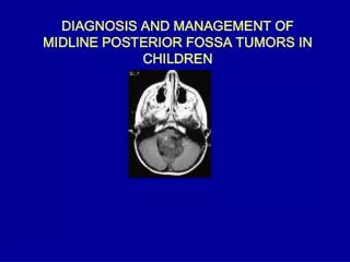
Computedtomography Contrast-enhanced CT scan of the abdomen reveals a bulky, circumscribed, lobulated, heterogeneous intra-abdominal mass measuring 18*13*17cm. Curvilinearfoci of highattenuation, consistent withcalcification, delimittumor lobules. The origin of this mass were impossible to determine.
Biology: plasma cortisol , testosterone αFP, βHCG, CA125 normal • Biopsy+ histologicalexamination: adrenocorticalcarcinoma
* Metastasis: • Pulmonarymetastasis
Treatment: • chemotherapywasproposed to the parents howevertheyrefused all treatment.
DISCUSSION • Adrenocorticalcarcinoma (ACC) comprises only 0.002% of all childhood malignancies and is potentially lethal. • Occur more frequently between the age of 3 and 5. • These hormone-secretingneoplasms are manifested by virilization, Cushing’s Syndrome, aldosteronism, or feminization.
Occur more frequently between the age of 3 and 5. • It’s now recongnized that most children with an adrenocortical neoplasm show clinical evidence of an endocrine abnormality, in contrast to the behaviour of adrenocortical tumors in adults. • These hormone-secretingneoplasms are manifested by virilization, Cushing’s Syndrome, aldosteronism, or feminization
Virilizationsignsinclude an increase in muscular mass, rapid growth, acne, pubic and facial hair, hirsutism, and an increase in the size of the penis or clitoris. these signs appeared in our patient
Children Typicallyguided by clinicalpresentation Adults Incidentallydiscovered in asymptomaticadults Diagnosisimaging of adrenocorticalneoplasms
Predisposingconstitutionalgeneticfactorshave been found in approximately 50% of childrenwith ACC. Twogenetic syndromes are clearly associated with ACC: • Beckwith-Wiedemann syndrome:alteration of the 11p15 region. • Li-Fraumeni syndrome:alterations of the tumor suppressor gene p53 on chromosome 17p • In familieswith the Li-Fraumenisyndromthe frequency of adrenocortical tumors is 100 times that in the general population.
Metastasis: • The lungis the mostcommon site of metastasisfollowed in frequency by the liver. • Othermetastatic sites: * The peritoneum ( 29%) * pleura or diaphragm (24%) * abdominal lymphnodes (24%) * Kidney ( 18%) * Venousextention * Cerebralmetastasis are extremely rare +++
Cross-sectionalimagingstudiesincluding US, CT, and magneticresonnance (MR) imaging, have largelysuppleated use of invasive procedures: • US and CT : principal diagnosismodalitiesused. • Theyare useful to suggest the diagnosis, and define local and distant extension as well as they guide biopsy.
Cross-sectional imaging studies typically demonstrate a large, circumscribed, predominantly solid suprarenal mass with variable heterogeneity due to hemorrhage and necrosis. Calcification is not uncommon. Local invasion and metastases to the lungs, liver, and regional lymph nodes may be present at diagnosis. When friable tumor thrombus extends into the inferior vena cava, it poses a high risk of pulmonary embolization.
Surgery is the only mode of therapy documented as effective for treating paediatric ACT • Radiotherapy has not yielded good results. • The role of chemotherapy has not been systematically evaluated in childhood ACC. • Currently, ortho-para-DDD, also known as MITITANE, is the chemotherapeutic agent used to treat ACC in adults. • Little information is available about the use of mitotanein children but response rates appear to be similar to those seen in adults with ACT
CONCLUSION This observation underscore: • Imaging importance in diagnosing and evaluating the extension of these tumors. • The importance of considering genetic testing and counselling for families of young children with ACC.
BIBLIOGRAPHY • 1. Sandrini R, Ribeiro RC, DeLacerda L, 1997 Childhoodadrenocorticaltumors. J Clin EndocrinolMetab 82: 2027-2031. 2. Wolthers OD, Cameron FJSI, Honour JW, Hindmarsh PC, Savage MO, Stanhope RGBCGD, 1999 Androgensecretingadrenocorticaltumours. Arch Dis Child 80: 46-50. 3. Ribeiro RC, Michalkiewicz EL, Figueiredo BC, et al, 2000 Adrenocorticaltumors in children. Braz J Med BiolRes 33: 1225-1234. 4. Michalkiewicz E, Sandrini R, Figueiredo B, et al, 2004 Clinical and outcomecharacteristics of childrenwithadrenocorticaltumors: a report from the International PediatricAdrenocorticalTumorRegistry. J Clin Oncol 22: 838-45. 5. Hanna AM, Pham TH, Askegard-Giesmann JR, et al, 2008 Outcome of adrenocorticaltumors in children. J PediatrSurg 43: 843-849. 6. Visser HK, 1966 The adrenal cortex in childhood. 2. Pathological aspects. Arch Dis Child 41: 113-136. 7. Stratakis CA, 2008 Cushing syndrome caused by adrenocorticaltumors and hyperplasias (corticotropin- independent Cushing syndrome). EndocrDev 13: 117-132. 8. Magiakou MA, Mastorakos G, Oldfield EH, et al, 1994 Cushing’s syndrome in children and adolescents. Presentation, diagnosis, and therapy. N Engl J Med 331: 629-636.9.Ciftci AO, Senocak ME, Tanyel FC, Buyukpamukcu N, 2001 Adrenocorticaltumors in children. J PediatrSurg 36: 549-554.