Download
1 / 135
1.35k likes | 1.54k Views
Unit 4: Cytology. The Study of the cell Cell: the smallest unit that can carry on all of life’s processes. EARLY MICROSCOPES. Zacharias Janssen - made 1st compound microscope a Dutch maker of reading glasses (late 1500’s ). Leeuwenhoek. The first to observe living cells ( 1675)

E N D
Unit 4: Cytology The Study of the cellCell: the smallest unit that can carry on all of life’s processes
EARLY MICROSCOPES • Zacharias Janssen - made 1st compound microscope • a Dutch maker of reading glasses (late 1500’s)
Leeuwenhoek • The first to observe living cells ( 1675) • discovered blood cells, bacteria and other single-celled organisms which he named “animacules
Leeuwenhoek’s Microscope • made a simple microscope (mid 1600’s) • magnified 270X • Early microscope lenses made images larger but the image was not clear
Leeuwenhoek's microscope A) a screw for adjusting the height of the object being examined B) a metal plate serving as the body C) a skewer to impale the object and rotate it D) the lens itself, which was spherical
Discovery of cells • Cells were first seen in 1665 by the early microscopist Robert Hooke. • Hooke was examining cork wood, and noticed that the wood was divided into little squares or “cells”
CELL THEORY • A theory resulting from many scientists’ observations & conclusions
CELL THEORY1. The basic unit of life is thecell. (Hooke) • In 1665, an English scientist named Robert Hooke made an improved microscope and viewed thin slices of cork viewing plant cell walls • Hooke named what he saw "cells"
CELL THEORY2. All living things are made of1 or more cells. • Matthias Schleiden(botanist studying plants) • Theodore Schwann(zoologist studying animals) stated that all living things were made of cells Schwann Schleiden
CELL THEORY3. All cells divide & come from oldcells. (Virchow) Virchow
MODERN MICROSCOPES • A microscope is simple or compound depending on how many lenses it contains • A lens makes an enlarged image & directs light towards you eye
A simple microscope has one lens • Similar to a magnifying glass • Magnificationis the change in apparent size produced by a microscope
COMPOUND MICROSCOPE • A compound microscope has multiple lenses • (eyepiece & objective lenses)
STEREOMICROSCOPE • creates a 3D image
ELECTRON MICROSCOPES • More powerful; some can magnify up to 1,000,000X • Use a magnetic field in a vacuum to bend beams of electrons • Images must be photographed or produced electronically
ScanningElectron Microscope (SEM) Electron microscope image of a spider Electron microscope image of a fly foot • produces realistic 3D image • only the surface of specimen can be observed
TransmissionElectron Microscope (TEM) • produces 2D image of thinly sliced specimen • detailed cell parts (only inside a cell) can be observed
ScanningTunneling Microscope (STM) • able to show arrangement of atoms
Ocular Lens Body Tube Nose Piece Arm Objective Lenses Stage Stage Clips Coarse Adj. Diaphragm Fine Adjustment Light Source Base Skip to Magnification Section
Magnification • To determine your magnification…you just multiply the ocular lens by the objective lens • Ocular 10x Objective 40x:10 x 40 = 400 So the object is 400 times “larger” Objective Lens have their magnification written on them. Ocular lenses usually magnifies by 10x
TOTAL MAGNIFICATION • Powers of theeyepiece (10X) multiplied by objective lenses determine total magnification.
Using a Microscope • Start on the lowest magnification • Don’t use the coarse adjustment knob on high magnification…you’ll break the slide!!! • Place slide on stage and lock clips • Adjust light source (if it’s a mirror…don’t stand in front of it!) • Use fine adjustment to focus
FROM CELL TO ORGANISM Tissue Group of cells working together Organ Group of tissues working together Organ System Group of organs working together Organism Any livingthing made of 1 or more cells
Two basic cell types: Eukaryotes (Eu = true) (kary = nucleus) Organisms whose cells contain a membrane-bound nucleus and other organelles. Prokaryotes (Pro = before) Organisms without a membrane-bound nucleus (bacteria). * These cells have genetic information, but not in a nucleus. * Evolutionists chose the prefix “pro” because they believe these evolved before others.
Prokaryotic Cells: Organisms with prokaryotic cells are called “prokaryotes” Prokaryotes have no true nucleus or organelles. Have a single strand of “looped” DNA Most prokaryotes are single-celled microscopic organisms.
Some Example Prokaryotes Coccus-shaped bacterium Bacillus-shaped bacterium Spirillum-shaped bacterium
Eukaryotic Cells: • Organisms composed of eukaryotic cells are called “eukaryotes.” • Have a membrane bound nucleus which contains the cell’s DNA. • Some eukaryotes are one-celled organisms.ALL multicellular organisms are eukaryotes. • Have organelles, each of which is surrounded by (or bound in) a “plasma membrane.”
Some Example Eukaryotes Plant leaf cells Yeast cells Fertilized human egg cell Nerve cells
Very simple cells(no organelles) Always single-celled No nucleus DNA arranged in one single loop Found only in kingdom Monera (bacteria) Complex cells( have organelles) Can be single-celled or multicellular Have a nucleus DNA arranged in many separate strands Found in Animal, Plant, Protists, and Fungi kingdoms Prokaryotes vs. Eukaryotes
Bacteria Eukaryotic cell 1.0-10.0 micrometers 10.0-100.0 micrometers Cell Size Type Diameter Cells range in size from 1mm ( frog egg) to 1micrometer
Prokaryotic bacteria cells surrounding a eukaryotic cell (possibly a white blood cell?)
Cell Diversity - Size • - • Size is limited by ratio between outer surface area and inner volume. • (Volume increases with the cube of the side length) • (Surface area increases by the square of the side length) • - So, as it grows, the surface area is too small to allow enough materials to pass through the membrane (water, food, waste).
Remember cells are not really cubes or even perfect spheres. We use cubes only as examples. • When a side of a cube equals "s", then the area of only one face is (A = s2). • The total surface area (T.S.A.) of a cubed cell is (T.S.A. = 6 x s2). • The volume of a cubed cell is (V = s3). • The distance from the center of the cell to each wall is (Distance = s divided by 2). ** Be able to calculate T.S.A., V, and distance if given “s” for a cell. ** Note the smallest cell has the largest T.S.A. to V ratio, and the smallest distance from center to membrane.
EXAMPLES: Nerve cells (for many connections), skin (for a flat covering), white blood cells (to travel & clean up) Cell Diversity - Shape Diversity of forms reflects diversity of function.
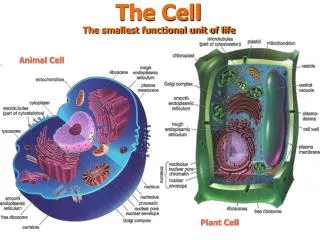
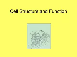
The 3 basic structures found in most cells: 1. Nucleus 2. Cytoplasm 3. Cell membrane
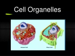
1. No cell wall 2. No chloroplast 3. No vacuole 4. shape is different 1. have cell wall 2. have chloroplast 3. have vacuole Between the two Animal Vs Plant Cell Animal Plant
Eukaryotic Cellhttp://www-class.unl.edu/bios201a/spring97/group6/eukcell.jpg
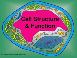
Ch 4.2 - Parts of Eukaryotic Cells Vesicles Endoplasmic Reticulum Nucleolus Golgi Complex Cytoplasm Ribosomes DNA Nucleus Cell Membrane Mitochondria
Internal Organization: • Organelles = perform specific functions. • function like tiny organs, analogous to organs of a multicellular body. • Cell Membrane = surrounds, contains, and protects the cell • Nucleus = large organelle containing most of the genetic information
CELL WALL • protects the cell • gives shape • is made of cellulose • A cell wall is found in plants, algae, fungi, & most bacteria.
CELL MEMBRANE (Plasma membrane) • Outer covering, protective layer around ALL cells • For cells with cell walls,the cell membrane is inside the cell wall • Allows food, oxygen, & water into the cell & waste products out of the cell.
CELL MEMBRANE (Plasma membrane) • The boundary of the cell…separates inside from outside of cell • Is Semipermeable Membrane: allows some substances into cell and keeps others out of cell.
CELL MEMBRANE (Plasma membrane) • Has a phospholipid bilayer. The lipid molecules are fluid and can move past one another in a fluid manner…also allows proteins to move and change in this layer thus scientist explain cell membrane and call it a Fluid Mosaic Model
CYTOPLASM • gelatin-like inside cell membrane • constantly flows • aka protoplasm • It contains the various organelles of the cell