Download
1 / 28
280 likes | 444 Views
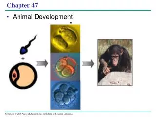
Chapter 47. Animal Development. Development is determined by the zygote’s genome and differences between embryonic cells Cell differentiation is the specialization of cells in structure and function

E N D
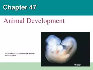
Chapter 47 Animal Development
Development is determined by the zygote’s genome and differences between embryonic cells • Cell differentiation is the specialization of cells in structure and function • Cytoplasmic determinants, the uneven distribution of maternal substances within the unfertilized egg, cause early embryonic cells to differentiate in some species. • Morphogenesis is the process by which an animal takes shape
Concept 47.1: After fertilization, embryonic development proceeds through cleavage, gastrulation, and organogenesis • Important events regulating development occur during fertilization and the three stages that build the animal’s body • Cleavage: cell division creates a hollow ball of cells called the blastula. • Gastrulation: produces a 3-layered embryo called the gastrula. • Organogenesis: generates rudimentary organs from which adult structures grow.
Fertilization • Fertilization brings the haploid (1 set of chromosome) nuclei of sperm and egg together, forming a diploid (2 sets of chromosomes) zygote • The sperm’s contact with the egg’s surface initiates metabolic reactions in the egg that trigger the onset of embryonic development (“activates the egg”)
LE 47-3 Contact and fusion of sperm and egg membranes Entry of sperm nucleus Acrosomal reaction Sperm plasma membrane Sperm nucleus Cortical reaction Contact Acrosomal process Basal body (centriole) Sperm head Fertilization envelope Fused plasma membranes Cortical granule Actin Perivitelline space Hydrolytic enzymes Acrosome Jelly coat Cortical granule membrane Vitelline layer Sperm-binding receptors Egg plasma membrane EGG CYTOPLASM
The Acrosomal Reaction • The acrosomal reaction is triggered when the sperm meets the egg. • This process begins when a specialized vesicle at the tip of the sperm called the acrosome, discharges hydrolytic enzymes. • This reaction releases hydrolytic enzymes that digest material surrounding the egg • This allows the acrosomal process (elongated sperm structure) to penetrate the jelly coat.
Molecules of a protein on the tip of the acrosomal process adhere to molecules of a specific receptor proteins on the egg’s surface. • This gamete contact and/or fusion depolarizes the egg cell membrane and sets up a fast block to polyspermy (multiple sperm). • Depolarization occurs when the ion channels open on the egg’s plasma membrane allowing sodium ions to flow into the egg cell and change the membrane potential. • Depolarization occurs within 1-3 seconds after a sperm binds to an egg.
Fusion of egg and sperm also initiates the cortical reaction • This reaction induces a rise in Ca2+from the egg’s ER into the egg’s cytosol. This causes cortical granules in the egg to fuse with the plasma membrane and discharge their contents. This leads to swelling of the perivitelline space, hardening of the vitelline layer, and clippling of sperm binding receptors. • These changes cause formation of a fertilization envelope that functions as a longer-term slow block to polyspermy. • Does occur in vertebrates (fishes and animals)
Activation of the Egg • The sharp rise in Ca2+ in the egg’s cytosol increases the rates of cellular respiration and protein synthesis by the egg cell • With these rapid changes in metabolism, the egg is said to be activated • Sperm cells do not contribute any materials required for activation. The unfertilized eggs of many species can be artificially activated by the injection of Ca2+ or by a variety of mildly injurious treatments, such as temperature shock.
LE 47-5 1 Binding of sperm to egg Acrosomal reaction: plasma membrane depolarization (fast block to polyspermy) 2 3 4 6 Seconds 8 10 Increased intracellular calcium level 20 Cortical reaction begins (slow block to polyspermy) 30 40 50 Formation of fertilization envelope complete 1 2 Increased intracellular pH 3 4 Increased protein synthesis 5 Minutes 10 20 Fusion of egg and sperm nuclei complete 30 Onset of DNA synthesis 40 60 First cell division 90
Fertilization in Mammals • Fertilization in other species share the same timing as the sea urchin in the previous slide. However, timing differs with species. • Sea urchins meiosis is already completed when the egg is released from the female. • In humans, the unfertilized egg stays at metaphase of meiosis II. Meiosis is NOT completed until they are fertilized in the female reproductive tract. • Fertilization is generally internal.
Fertilization in Mammals continued • Secretions in the mammalian female trace alter certain molecules on the surface of sperm cells and also increase sperm motility. • The mammalian egg is cloaked by follicle cells released along with the egg during ovulation. The sperm must migrated through this layer of follicle cells before it reaches the zona pellucida. • In mammalian fertilization, the cortical reaction modifies the zona pellucida as a slow block to polyspermy
3. Breakdown of the zona pellucida by these enzymes allows the sperm to reach the plasma membrane of the egg. Membrane proteins of the sperm bind to the receptors on the egg membrane, and the two membranes fuse LE 47-6 4. The nucleus and other components of the sperm cell enter the egg. 2. This binding induces the acrosomal reaction, in which the sperm released hydrolytic enzymes into the zona pellucida. 1. Sperm migrated through the coat of follicle cells and binds to receptor molecules in the zona pellucida of the egg. Follicle cell Sperm basal body Cortical ganules Zona pellucida Sperm nucleus Egg plasma membrane 5. Enzymes released during the cortical reaction harden the zona pellucida, which now functions as a block to polyspermy. Acrosomal vesicle EGG CYTOPLASM
Fertilization in Mammals continued • After the egg and sperm membrane fuse, the whole sperm, tail and all is taken into the egg. • The egg lacks a centrosome. The basal body of the sperm’s flagella now acts as the centrosome and wraps itself around the centriole. • This will allows mitotic spindles to form for the first cell division. • Fertilization is much slower in mammals. The first cell division occurs 12-36 hours after sperm binding in mammals.
Cleavage cell division creates a hollow ball of cells called the blastula. • Fertilization is followed by cleavage, a period of rapid cell division without growth • Cells undergo S and M phases of the cell cycle but skip Gap 1 and Gap 2. Little or now protein synthesis occurs. • The embryo does not enlarge during this period of development. • Cleavage partitions the cytoplasm of one large cell into many smaller cells called blastomeres. Each with its own nucleus.
LE 47-7 Fertilized egg Four-cell stage Morula Blastula • First 5-7 divisions from a cluster of cells known as the morula • A fluid-filled cavity called the blastocoel begins to form within the morula and is fully formed in the blastula, a hollow ball of cells. • During cleavage, different regions of the cytoplasm end up in separate blastomeres. These regions may contain different cytoplasmic determinants, in many species this partitioning sets the stage for subsequent developmental events.
Gastrulation • Gastrulation rearranges the cells of a blastula into a three-layered embryo, called a gastrula, which has a primitive gut. • Varies from one animal to another, the process is driven by change in cell motility, changes in cell shape, and changes in cellular adhesion to other cells and to molecules of the extracellular matrix. • This results in the three cell layers.
The three layers produced by gastrulation are called embryonic germ layers • The ectoderm forms the outer layer • The endoderm lines the digestive tract • The mesoderm partly fills the space between the endoderm and ectoderm • Eventually, these three cell layers develop into all the tissues and organs of the adult animal. Video: Sea Urchin Embryonic Development
Organogenesis • During organogenesis, various regions of the germ layers develop into rudimentary organs • Early in vertebrate organogenesis, the notochord forms from mesoderm, and the neural plate forms from ectoderm
Mesoderm lateral to the notochord forms blocks called somites • Lateral to the somites, the mesoderm splits to form the coelom • The neural plate soon curves inward, forming the neural tube • Many structures are derived from the three embryonic germ layers during organogenesis
Developmental Adaptations of Amniotes • Because all vertebrate embryos required an aqueous environment for development, embryos of birds, other reptiles, and mammals develop in a fluid-filled sac in a shell (birds & reptiles) or the uterus (marsupials & eutherian) • Organisms with these adaptations are called amniotes • In these organisms, the three germ layers also give rise to the four membranes that surround the embryo
LE 47-17 Amnion Allantois Embryo Amniotic cavity with amniotic fluid Albumen Shell Yolk (nutrients) Yolk sac Chorion
Mammalian Development • Fertilization takes place in the oviduct, and the progresses as the embryo completes its journey down the oviduct to the uterus. • The eggs of placental mammals • Are small and store few nutrients • Exhibit holoblastic cleavage (complete cell division of egg, having little or moderate amount of yolk) • Gastrulation and organogenesis resemble the processes in birds and other reptiles • Early cleavage is relatively slow in humans and other mammals
At completion of cleavage, the blastocyst forms • The trophoblast, the outer epithelium of the blastocyst, initiates implantation in the uterus, and the blastocyst forms a flat disk of cells • As implantation is completed, gastrulation begins • The extraembryonic membranes begin to form • By the end of gastrulation, the embryonic germ layers have formed
LE 47-18a Endometrium (uterine lining) Inner cell mass Trophoblast Blastocoel Blastocyst reaches uterus. Expanding region of trophoblast Maternal blood vessel Epiblast Hypoblast Trophoblast Blastocyst implants.
LE 47-18b Expanding region of trophoblast Amniotic cavity Amnion Epiblast Hypoblast Chorion (from trophoblast Yolk sac (from hypoblast) Extraembryonic membranes start to form and gastrulation begins. Extraembryonic mesoderm cells (from epiblast) Allantois Amnion Chorion Ectoderm Mesoderm Endoderm Yolk sac Extraembryonic mesoderm Gastrulation has produced a three-layered embryo with four extraembryonic membranes.
The extraembryonic membranes in mammals are homologous to those of birds and other reptiles and develop in a similar way