Download
1 / 26
260 likes | 321 Views
Acetylcholine Action potential Amygdale Autonomic nervous system Axon Basal ganglia Biogenic amines Biological clock Blood-brain barrier Brain Brainstem Cell body Central canal Central nervous system (CNS) Centralization Cephalization Cerebellum Cerebral cortex

E N D
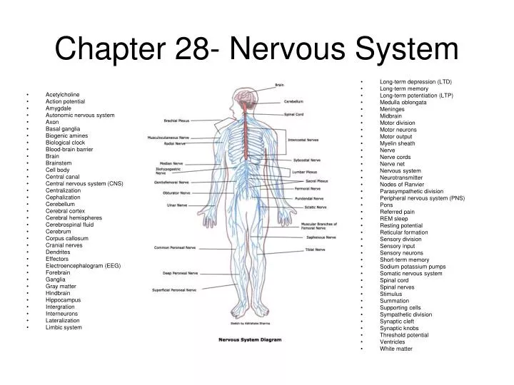
Acetylcholine Action potential Amygdale Autonomic nervous system Axon Basal ganglia Biogenic amines Biological clock Blood-brain barrier Brain Brainstem Cell body Central canal Central nervous system (CNS) Centralization Cephalization Cerebellum Cerebral cortex Cerebral hemispheres Cerebrospinal fluid Cerebrum Corpus callosum Cranial nerves Dendrites Effectors Electroencephalogram (EEG) Forebrain Ganglia Gray matter Hindbrain Hippocampus Intergration Interneurons Lateralization Limbic system Long-term depression (LTD) Long-term memory Long-term potentiation (LTP) Medulla oblongata Meninges Midbrain Motor division Motor neurons Motor output Myelin sheath Nerve Nerve cords Nerve net Nervous system Neurotransmitter Nodes of Ranvier Parasympathetic division Peripheral nervous system (PNS) Pons Referred pain REM sleep Resting potential Reticular formation Sensory division Sensory input Sensory neurons Short-term memory Sodium potassium pumps Somatic nervous system Spinal cord Spinal nerves Stimulus Summation Supporting cells Sympathetic division Synaptic cleft Synaptic knobs Threshold potential Ventricles White matter Chapter 28- Nervous System
The nervous system • Receives signals, interprets them and sends out appropriate commands • 2 divisions: • Central- CNS- brain and spinal cord- where integration occurs • Peripheral- PNS- nerves- carry signals in and out of CNS
The neuron • Nerve cell- carries the signal • 3 functions: • Sensory input: signal from sense receptor to integration center • Integration: interpretation of sensory signals and formation of responses • Motor output: conduction of signal from integration center to effector- which performs body’s response • Types of neurons- • Sensory- convey info from sense receptor to CNS • Interneuron- relays signal to other interneurons or motor neurons • Motor- CNS to effectors
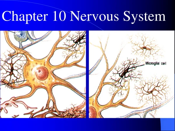
Parts of a neuron • Cell body- holds nucleus and organelles • Dendrites- short branches off cell body, receives messages • Axon- longer, usually 1 fiber, sends signal towards other neuron or effector • Supporting cells- protect, insulate and reinforce neurons • Myelin sheath- made of Schwann cells- insulates • Nodes of Ranvier- space between Schwann cells, signal jumps from node to node to move faster
Nerve- cable-like bundle of neuron extensions wrapped in CT • Ganglia- clusters of neuron cell bodies in the nerves
Nerve signal • Change in membrane potential • Resting potential- voltage across the plasma membrane • Diffusion of ions and Na/K pumps maintain resting potential
Nerve signal con’t • Stimulus- factor that causes a nerve cell to be generated • Action potential- nerve signal- change in the membrane voltage • When stimulus is applied- threshold voltage is reached • Change in charge is caused by rapid movements of Na and K at membrane channels
Nerve signal con’t • Action potential propagates itself • Electrical changes in 1 section trigger them in another • Frequency of action potentials changes with intensity of stimulus
Synapse • Space between 2 neurons or a neuron and an effector cell • Signal sent can be electrical or chemical • Synaptic cleft- gap between neurons, prevents action potential from sending info, action potentials can be converted to chemical signals (neurotransmitters) • The action potential triggers vesicles to fuse with plasma membrane • Neurotransmitters bind to receptors and open ion channels to ions that start new action potential or stops one • Neurotransmitter is then broken down or taken back into signaling cell
Fig 28.7 • Excitatory and inhibitory signals
Types of neurotransmitters • Most- small N containing organic molecules • Acetylcholine- used in brain and between motor neurons and muscle cells • Biogenic amines- important in CNS • Many drugs act out chemical signals • Caffeine- countering effects of inhibitory neurotransmitters =awake • Nicotine- acts as a stimulant by binding to and activating acetylcholine receptors • Alcohol- depressant, inhibits effects of certain neurotransmitters • Prozac- antidepressant, blocks removal of serotonin increasing amount of mood altering neurotransmitters available • Amphetamines & cocaine- stimulants, increase in release and availability of norepinephrine and dopamine
Nervous system organization correlates with body symmetry • Hydra-nerve net • Cephalization- concentration of NS in head end • Centralization- presence of CNS
Parts of our nervous system • CNS • Spinal cord- inside vertebral column- where reflexes are controlled • Blood-brain barrier- capillaries in brain restrict many substances from entering • Brain and SC- filled with cerebrospinal fluid- cushions CNS, supplies nutrients, hormones WBC’s • Meninges- layers of CT that protect CNS • White matter- mainly axons (myelin sheaths) • Gray matter- mainly nerve bodies and dendrites • Cranial nerves- signal to and from brain • Spinal nerves- 2 and from spine
Parts of our nervous system • PNS (2 divisions) • Sensory- senses internal and external env • Motor – (2 divisions) • Somatic- voluntary • Autonomic- involuntary- regulates internal environment • Parasympathetic- primes body for digestion and rest- activities that gain/conserve E • Sympathetic- prepares body for intense E- E consuming activities
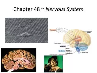
The brain • Hindbrain and midbrain- • Pons, medulla oblongata and midbrain- brainstem- sensory filter, regulates sleep, coordinates body movement • Pons and medulla oblongata- control breathing, and med. Obl. also controls circulation, swallowing, digestion • Cerebellum- planning center for body movements, learning and remembering
The Brain • Forebrain • Thalamus- relay info to cerebral cortex • Sorts data as to what goes to cerebrum and what comes out • Hypothalamus- regulates basics (body temp, BP, thirst, hunger) and biological clock- maintains daily biorhythms • Cerebrum- consists of hemispheres- site of memory, learning, emotion, speech, formulate complex behavior • Left side of cerebrum- language, logic, math • Right side- spatial, pattern, face recognition, music, emotional processing
Limbic system • Emotion, learning and memory • Includes parts of thalamus, hypothalamus, amygdala and hippocampus • Amygdala- recognize emotion in facial expression and laying down emotional memories • Hippocampus- involved in formation of memories • Moving from short to long term- enhanced by rehearsal, positive or negative emotional states and association to previous learned data