Download
1 / 49
530 likes | 802 Views
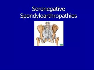
Spondyloarthropathies John Imboden MD. 23 y.o. woman with low back pain. 9 years of low back pain Spontaneous, insidious onset at age 14 persistent, dull, non-radiating improved by mild-moderate activity made worse by inactivity associated with AM stiffness for >3 hours

E N D
23 y.o. woman with low back pain • 9 years of low back pain • Spontaneous, insidious onset at age 14 • persistent, dull, non-radiating • improved by mild-moderate activity • made worse by inactivity • associated with AM stiffness for >3 hours • Episode of “eye inflammation” age 12 • Family History: Unremarkable • Social History: Full-time college student
23 y.o. woman with low back pain • On examination: decreased range of motion of her lumbar spine and decreased chest expansion • She has had some relief with NSAIDs but now her back pain is so severe she has had to cut back on her course load and is uncertain whether she can continue college. • She has seen multiple physicians in the past 9 years but none has been able to make a diagnosis. • What is the likely cause of the back pain?
Spondyloarthropathies • Ankylosing spondylitis • Reactive arthritis • Arthritis associated with inflammatory bowel disease (Crohn’s disease and ulcerative colitis) • Arthritis associated with psoriasis • Undifferentiated spondyloarthropathy
Spondyloarthropathies: common features • Involvement of the axial skeleton • Asymmetric oligoarthritis of peripheral joints • Enthesitis & dactylitis • Seronegative • negative tests for rheumatoid factor and ANA • Association with HLA-B27
Involvement of axial skeleton:sacroiliac joints and all components of spine Sacroiliitis occurs in 100% cases of ankylosing spondylitis
Peripheral arthritis: asymmetric oligoarthritis with predilection for large joints of lower extremities Peripheral arthritis occurs in the great majority of patients with reactive arthritis or psoriatic arthritis but in <25% with ankylosing spondylitis
Enthesitis inflammation and boney proliferation where plantar fascia inserts onto the calcaneus
Enthesitis: inflammation where tendon, ligament, or joint capsule attach to bone Spondyloarthopathy: - Enthesitis - Synovitis T cell and macrophage Infiltration Local cytokine production: IL-1,IL-6, IL-17, TNF-a
HLA-B27 in Caucasian populations in US normal controls 8% ankylosing spondylitis 90% reactive arthritis with spondylitis 60-80% psoriatic with spondylitis 50% IBD with spondylitis 50% HLA-B27 and spondyloarthropathies
HLA polymorphisms and selected autoimmune diseases DiseaseHLA markerrelative risk ankylosing spondylitis B27 90 reactive arthritis B27 40 rheumatoid arthritis DR4 5
HLA-B27 and risk of spondyloarthropathy • Strongest association between an HLA gene and a rheumatic disease BUT • HLA-B27 not absolutely required • HLA-B27 not sufficient • <20% of B27+ individuals develop disease
HLA-B27 and risk of spondyloarthropathy Environmental triggers mucosal inflammation (infection, IBD) psoriasis unknown + Spondyloarthropathy Genetic background HLA-B27 other genes
Ankylosing Spondylitis • An inflammatory arthritis with predilection for the axial skeleton • Sacroiliac joints • always involved • bilateral • early in the course of the disease • Spine (cervical, thoracic, lumbar) • variable in severity and extent
Ankylosing spondylitis: a genetically determined disease • Family and twin studies: largely a genetic disease • Multiple genes involved: HLA-B27 confers a relative risk of 90 but constitutes only 15-50% of the overall genetic risk • Environmental trigger is essential but ubiquitous
Ankylosing Spondylitis • male predominance: M:F, 3:1 • age of onset: 15 to 35 years - rarely begins after age 50 • usual presenting complaint: low back pain
Age of onset of symptoms in AS(Feldkeller et al. Rheumatol Int 23: 61, 2003
Ankylosing spondylitis: “inflammatory” back pain • insidious in onset • persistent • dull in quality • associated with stiffness • worse in AM or after prolonged inactivity • eased by mild activity
Physical examination in ankylosing spondylitis • Tenderness over the sacroiliac joints • Limited range of motion of the spine • Decreased chest expansion • due to inflammation of the costovertebral joints
Ankylosing spondylitis Involvement of cervical spine: Inability to touch occiput to wall Involvement of lumbar spine: Failure to reverse lumbar lordosis during flexion
Progression of ankylosing spondylitis: lumbar spine facet “squared-off” syndesmophytes disease vertebrae
Ankylosing spondylitis:syndesmophytes and fusion of lumbar spine • Spinal complications of AS: • loss of motion • osteopenia • increased risk of fracture • - C1-C2 subluxation
Ankylosing spondylitis spine fused in flexion
Ankylosing spondylitis: extraarticular manifestations • common: - anterior uveitis (20-40%) • Associated with HLA-B27 - GI inflammation (subclinical) • uncommon/rare: - aortitis (3% after 15 years) - apical fibrosis of the lung
Ankylosing spondylitis: diagnosis • Diagnosis is best secured by combination of inflammatory low back pain plus radiographic evidence of sacroiliitis • But plain radiographs may fail to reveal changes for years • MRI of SI joints • HLA-B27 testing • Average delay in diagnosis: 8 years
23 y.o. woman with low back pain Key features: • Age of onset: 14 • Quality of the back pain: inflammatory • Past history of ocular inflammation • Decreased L spine motion and chest expansion
23 y.o. woman with low back pain • Radiograph: bilateral sacroiliitis • Diagnosis: ankylosing spondylitis • Started on anti-TNF therapy • AM stiffness 180 min 0 min • Chest expansion 2 cm 4.5 cm • Returned to college full time
Ankylosing spondylitis: therapy • physical therapy to maintain erect posture • NSAIDs • avoid use of systemic corticosteroids • Anti-tumor necrosis factor therapy • Symptomatic improvement in axial skeleton disease
Reactive arthritis • arthritis triggered by GU or GI infections in which the inciting organism cannot be cultured from involved joints • genitourinary infections: - Chlamydia trachomatis • enteric infections: - Shigella - Salmonella - Yersinia - Campylobacter
Reactive Arthritis chronic arthritis GI/GU 1 -4 wks reactive infection arthritis (1-4%) months can be “idiopathic”resolution
Reactive Arthritis • Cultures of synovial fluid and synovial tissue are sterile • Bacterial antigens can be detected in synovial tissue, even years after the onset of arthritis • No evidence of viable organisms • Antibiotics: • No proven benefit for enteric forms
Reactive arthritis: general features • M:F, 5:1 • often, at onset,constitutional symptoms with prominent weight loss, fatigue, & malaise • peripheral arthritis > axial arthritis • asymmetric oligoarthritis • lower extremity predominance • enthesitis (heel pain is common) • extraarticular disease
Reactive arthritis: extraarticular manifestations • eye: • conjunctivitis: usually mild • anterior uveitis • mucous membranes and skin: • urethritis • oral ulcers (painless) • keratoderma blenorrhagica • circinate balanitis • nail changes
Psoriatic arthritis • Peripheral arthritis • Develops in 5-7% of patients with psoriasis • Oligoarthritis, monarthritis • Polyarthritis • Arthritis mutilans • Spondylitis • Develops in 20% with peripheral arthritis
Psoriatic arthritis: DIP involvement Inflammation of DIP joint Nail pitting
Spondyloarthropathies: key points • Shared features • Why we group the spondyloarthropathies • How the spondyloarthropathies differ from RA • Major clinical manifestations of ankylosing spondylitis and reactive arthritis • Recognize clinical presentations of these diseases • Importance of HLA-B27 as a risk factor