Download

1 / 45
450 likes | 535 Views
Note: there are slides before these that we will take up. Neuron Membrane Potential. Muscle Membrane Potential. Muscle Tension. 1 2 3 4 5 6 7 8 9 10 96 97 98 99 100. Types of Muscle Contraction.

E N D
Neuron Membrane Potential Muscle Membrane Potential Muscle Tension 1 2 3 4 5 6 7 8 9 10 96 97 98 99 100
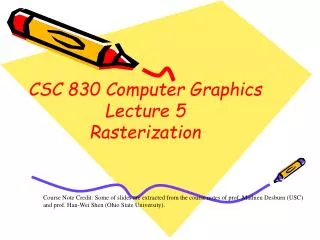
Types of Muscle Contraction Concentric Isometric Eccentric
Types of Muscle Contraction Force (g) 1.6 1.4 1.2 1.0 0.8 0.6 0.4 0.2 Maximal Isometric tension 0.8 0.6 0.4 0.2 0 0.2 0.4 0.6 0.8 Shortening(m/s) Concentric Lengthening (m/s) Eccentric Velocity
Functional Classification of Muscles • Agonists:prime movers; responsible for the movement • Antagonists:oppose the agonists to prevent overstretching of them • Synergists:assist the agonists and sometimes fine-tune the direction of movement
Functional Classification of Muscles In the control of muscular contraction, muscles rarely function in isolation
Control of Muscular Contraction • How is this all coordinated??? • How much force? • How fast? • Which muscles? • The answer relies on use of specific: • Motor Units • Fibre Types • Feedback Loops
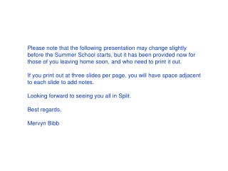
Muscle Fibre Types • 2 main categories: • Type I (Slow Twitch, Slow Oxidative, SO) • Type II (Fast Twitch) • More than one type of fast twitch fibre • Classified based on contractile speed and metabolic properties Type IIa(Fast Oxidative/Glycolytic, FOG, Fatigue Resistant) – slowest contractile speed of fast twitch fibres Type IId/x or IIb (Fast Glycolytic, FG, Fatigueable) – fastest contractile speed of fast twitch fibres (note: little Type IIbfound in humans)
Fibre Types Characteristics Type IIx/d 50 40 30 20 10 0 Tension Type IIa Type I 50 100 150 200 Time (msec) stimulus
Motor Units • Recall: All fibres of a motor unit are of the same type • It appears that the motor neuron determines fibre type • ST motor units are smaller (i.e. less fibres), therefore generate less force than FT motor units • Different muscles have different fibre compositions probably related to function
% ST Fibres Fibre Types Differ within Muscles For example: % ST in different muscles reflects function
Motor Units • Recall: different muscles have different fibre compositions probably related to function • Typically, successful athletes have fibre type profiles that may vary with event
$ 1,000,000 Question??? • It appears that the proportion of various fibre types can influence an athlete’s success, which begs the question…. … Is this a function of training or genetics???
% of Slow Twitch Fibres in Sets of Twins TWIN A 80 60 40 20 0 identical fraternal TWIN B 0 20 40 60 80
Resistance Training and Gains in Muscular Fitness • Muscle is very plastic (adaptable), increasing in size and strength with training and decreasing with immobilization • Strength is a function of muscle size. • Remember, more cross-bridges = more force.
Neural Control of Strength Gains All increases in strength possess some neural mechanisms: 1. Recruitment of motor units • Increased number of motor units recruited from increased neural drive • Synchronicity of motor unit recruitment is improved
Motor Unit Recruitment • Motor units are recruited in order of smallest to largest motor neuron – “Hennman’s Size Principle” • Slow twitch motor units, with smaller motor neurons, are recruited first in graded contractions • Fast twitch motor units are recruited as the force required increases
Neural Control of Strength Gains All increases in strength possess some neural mechanisms: 1. Recruitment of motor units • Increased number of motor units recruited from increased neural drive • Synchronicity of motor unit recruitment is improved 2. Increased frequency of discharge from the a-motor neuron
Neural Control of Strength Gains All increases in strength possess some neural mechanisms: 1. Recruitment of motor units • Increased number of motor units recruited from increased neural drive • Synchronicity of motor unit recruitment is improved 2. Increased frequency of discharge from the a-motor neuron 3. Decrease in autogenic inhibition 4. Reduction in the coactivation of agonist and antagonist muscles 5. Morphological changes in the neuromuscular junction
Neuromuscular Junction (NMJ) Fig 3.5
Long-term strength increases are largely the result of muscle fiber hypertrophy Muscle Hypertrophy Transient hypertrophy is the increase in muscle size that develops during and immediately following a single exercise bout Fluid accumulation in the interstitial and intracellular space from the blood plasma H H H H H H H H H H H H H H O O O O O O O
Long-term strength increases are largely the result of muscle fiber hypertrophy Muscle Hypertrophy Transient hypertrophy is the increase in muscle size that develops during and immediately following a single exercise bout Fluid accumulation in the interstitial and intracellular space from the blood plasma Chronic hypertrophy is the increase in muscle size after long-term resistance training Increased size: (Fiber Hypertrophy) Increased # muscle fibers (Fiber Hyperplasia)
Microscopic Views of Muscle Cross Sections Before and After Training Photos courtesy of Dr. Michael Deschene's laboratory.
Myonuclear Domains • Theory of myonuclear domains: • Each myonuclei supports a finite area of the muscle cell. In order for hypertrophy to occur, you need more nuclei!! • Where might they come from?
Fiber Hyperplasia • Muscle fibers can split in half with intense weight training (cat research) • Each half then increases to the size of the parent fiber • Conflicting study results may be due to differences in the training load or mode • Satellite cells may also be involved in the generation of new skeletal muscle fibers • Hyperplasia has been clearly shown to occur in animal models; only a few studies suggest this occurs in humans too
Muscle Atrophy and Decreased Strength Atrophy = decrease in size Immobilization, Disuse, Neural Disorders • Decreased rate of protein synthesis • Decreased strength • Decreased cross-sectional area • Decreased neuromuscular activity • Affects both type I and type II fibers, with a greater effect in type I fibers. WHY? • Muscles can recover when activity is resumed
Muscle Atrophy and Decreased Strength Cessation of Training • Decreased strength • Little change in fiber cross-sectional area (but … type II fiber areas tend to decrease) • Maintenance training is important to prevent strength losses
Muscle Soreness Acute Muscle Soreness • Felt during the later stages and immediately after exercise • Can result from accumulations of exercise end-products such as increase H+ and edema (fluid build up in the muscle)
Muscle Soreness Delayed-Onset Muscle Soreness (DOMS) • Felt in the days following heavy or unaccustomed exercise • Eccentric muscle action is the primary initiator of DOMS – not directly related to lactic acid (lactate)
DOMS • It is possible that the damage leading to DOMS is caused by excessive amounts of Ca2+ in the muscle cell – enters through tears in the sarcolemma • Ca2+ can activate proteases (e.g. Calpain) which can digest structural proteins • Ca2+ can also activate phosolipase which can lead to further damage of the lipid membrane
DOMS and Performance Loss in strength can be associated with: 1. Physical disruption • Z-line streaming, focal damage 2. Failure in ECC • Tears in sarcolemma. can hinder nerve impulse conduction • Triad disruption. 3. Loss of contractile proteins • Actin and myosin, as well as structural proteins
DOMS post-strenuous exercise pre-exercise • Muscle sample taken immediately after a marathon • Shows cell membrane (sarcolemma) disruption
DOMS post-strenuous exercise pre-exercise • Muscle sample taken immediately after a marathon • Shows Z-line streaming
DOMS and Performance • Force-generating capacity of injured muscles is decreased • Strength loss may persist for days or weeks
Recovery and Protection from DOMS • Once muscle damage and soreness has occurred, the muscle adapts such that it is protected against subsequent soreness • This adaptation can last 3-4 weeks, with some reporting as long as 6 months • This adaptation is associated with smaller losses in strength and decreased blood CK following exercise of the same intensity as the original damaging bout
Inflammation and Repair • Neutrophils (a type of white blood cell, WBC) enter the injured area and help to clean up the site of damage these release cytokines • By 10-15 days many fibres have become necrotic (dying) and mononucleated inflammatory cells become prominent • After 2-3 weeks, much of the damage has been repaired by regeneration of fibre segments – this involves satellite cells
Inflammation and Repair Control 1 Day 4 Days 3 Days 7 Days 14 Days
The Satellite Cell Responseto Muscle Injury Reprinted, by permission, from T.J. Hawke and D.J. Garry, 2001, “Myogenic satellite cells: Physiology to molecular biology,” Journal of Applied Physiology 91: 534-551.
Inflammation and Repair • Upon activation, satellite cells migrate to the necrotic area and differentiate into myoblasts • Similar to embryonic development, these myoblasts form myotubes (precursors to muscle cell formation) • As the myotubes extend, they reach the intact “stumps” of the damaged fibres and the fibres become continuous • The entire inflammation and repair process can take 30 days!