Download
1 / 39
440 likes | 812 Views
Joints & Articulations. Classification Of Joints. Classification By Function (degree of movement possible): Synarthroses (Syn=connected, immovable) Joints with little or no movement Skull sutures, cranium (minus the mandible ) Amphiarthroses (Amphi = on both sides, between)

E N D
Classification Of Joints • Classification By Function (degree of movement possible): • Synarthroses(Syn=connected, immovable) • Joints with little or no movement • Skull sutures, cranium (minus the mandible) • Amphiarthroses(Amphi = on both sides, between) • Slightly moveable joints • Intervertebral discs, costosternal joints, cartilaginous joints(vertebrate between spine) • Diarthroses(Diar=passing through, free moving) • Freely moveable joints • Shoulder, knee, hip, elbow, interphalangeal, tarsal, and carpal joints
Joint Classification • Classification by structure: • Synovial joints: • Bones separated by a joint cavity; lubricated by synovial fluid; enclosed in a fibrous joint capsule. • Shoulder, hip, elbow, knee, carpal, interphalangeal How would we classify these joints functionally?
Joint Classification • Fibrous joints: • Bones held together by collagenous fibers extending from the matrix of one bone into the matrix of the next. • No joint cavity • Skull sutures, teeth in joints, distal radioulnar joints & tibiofibular joints
Joint Classifications • Cartilaginous joints: • Bones held together by cartilage; no joint cavity • Epiphyseal plates of long bones, costosternal joints, pubic symphysis, intervertebral discs
Structure and Function Metopic Suture • Joints are designed for their function. • Let’s look at sutures as our 1st example: • Name 4 sutures! • What function do you suppose sutures are designed for? Saggital Suture Coronal Suture Coronal Suture Lambdoid Suture
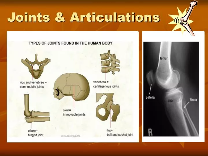
Types of Joints in the Human Body (1) Immovable: fixed joint such as the cranium (2) Ball-and-socket joints: such as the shoulder and hip joints, allow backward, forward, sideways, and rotating movements. (3) Hinge joints: such as in the fingers, knees, elbows, and toes, allow only bending and straightening movements. (4) Pivot joints: such as the neck joints, allow limited rotating movements. (5) Sliding Joint: found in the vertebral column and allows small sliding movements. The vertebrae have pads of cartilage between them, and the bones slide over these pads. This is what makes the backbone so flexible. (6) Ellipsoidal Joint: similar to a ball and socket joint. They allow the same type of movement to a lesser magnitude such as the wrist
Structure and Function • Now let’s talk about synovial joints. • 5 main structural characteristics: • Articular cartilage • What kind of cartilage is it? • Where do we find it? • What does it do? Hyaline
Structure and Function • Articular capsule • 2 layered. Surrounds both articular cartilages and the space btwn them. • External layer is made of dense irregular CT & is continuous w/ the perisoteum. • Inner layer is a synovial membrane made of loose connective tissue. • It covers all internal joint surfaces except for those areas covered by the articular cartilage.
Structure and Function • Joint(Synovial) Cavity • The potential space within the joint capsule and articular cartilage • Synovial Fluid • A small amount of slippery fluid occupying all free space w/i the joint capsule • Formed by filtration of blood flowing thru capillaries in the synovial membrane • Synovial fluid becomes less viscous as joint activity increases.
Structure and Function • ReinforcingLigaments • What kind of tissue are they? • What do you suppose their function is? • Double-jointed-ness results from extra-stretchy ligaments and joint capsules. Is this necessarily a good thing?
Other Synovial Structures • The knee and hip joints have cushioning fatty pads btwn the fibrous capsule and the synovial membrane or bone. • Discs of fibrocartilage (i.e., menisci) which improve the fit between bone ends, thus stabilizing the joint. • Found in the knee, jaw, and sternoclavicular joint. • Bursae are basically bags of lubricant - fibrous membrane bags filled with synovial fluid. Often found where bones, muscles, tendons, or ligaments rub together.
Types of Synovial Joints • Planejoints • Articular surfaces are flat and allow short slipping or gliding movements. • Intercarpal and intertarsal joints • Hinge joints • A cylindrical projection of one bone fits into a trough-shaped surface on another (like a hotdog in a bun) • Movement resembles a door hinge. • Elbow joint – ulna and humerus; Interphalangeal joints
Type of Synovial Joints • Pivot joints • Rounded end of one bone protrudes into a ring formed by another bone or by ligaments of that bone. • Proximal radioulnar joint • Atlas-axial joint • Condyloid joints • Oval articular surface of one bone fits into a complementary depression on another. • Radiocarpal joints • Metacarpophalangeal joints
Types of Synovial Joints • Saddle joints • Each articular surface has convex and concave areas. Each articular surface is saddle-shaped. • Carpometacarpal joints of the thumbs. • Ball-and-Socket joints • Spherical or semi-spherical head of one bone articulates with the cuplike socket of another. • Allow for much freedom of motion. • Shoulder and hip joints.
The Knee • Largest and most complex diarthrosis in the body. • Primarily a hinge joint, but when the knee is flexed, it is also capable of slight rotation and lateral gliding. • Actually consists of 3 joints: • Patellofemoral joint • Medial and lateral tibiofemoral joints • The joint cavity is only partially enclosed by a capsule – on the medial, lateral, and posterior sides.
The Knee • The lateral and medial condyles of the femur articulate with the lateral and medial condyles of the tibia. • Between these structures, we have the lateral and medial menisci. • Anteriorly, the patellar ligament binds the tibia (where?) to the inferior portion of the patella. The superior portion of the patella is then connected to the quadriceps femoris muscle
The Knee • At least a dozen bursae are associated with the knee. • Multiple ligaments are present. • The fibular collateral ligament extends from the lateral epicondyle of the femur to the head of the fibula. • The tibial collateral ligament connects medial epicondyle of the femur to the medial condyle of the tibial shaft and is also fused to the medial meniscus. • Both of these ligaments prevent excessive rotation
The Knee • The anterior and posterior cruciate ligaments are also very important. • ACL connects the anterior intercondylar area of the tibia to the medial side of the lateral femoral condyle. • Prevents forward sliding of the tibia and hyperextension of the knee. • PCL connects the posterior intercondylar area of the tibia to the lateral side of the medial femoral condyle. • Prevents backward displacement of the tibia or forward sliding of the femur.
ACLVideos Learn more about ACL Injury http://video.about.com/sportsmedicine/Anterior-Cruciate-Ligament.htm http://video.about.com/sportsmedicine/Medial-Meniscus-Injury.htm
Ankle Fracture Surgery Fibular Fracture
Clinical Conditions • Arthritis describes about 100 different types of inflammatory or degenerative joint diseases. • Osteoarthritis • Most common arthritis. • Normal joint use prompts the release of cartilage-damaging enzymes. If cartilage destruction exceeds cartilage replacement, we’re left with roughened, cracked, eroded cartilages. • Eventually bone tissue thickens and forms spurs that can restrict movement. • Most common in C and L spine, fingers, knuckles, knees, and hips.
Clinical Conditions • Rheumatoid arthritis • Chronic inflammatory disorder • Marked by flare-ups • Autoimmune disease. • Body creates antibodies which attack the joint surfaces • The synovial membrane can inflame and eventually thicken into a pannus – an abnormal tissue that clings to the articular cartilage.
Clinical Conditions • Gouty arthritis • When nucleic acids are metabolized uric acid is produced. Normally uric acid is excreted in the urine. • If blood [uric acid] rises due to decreased excretion or increased production, it may begin to form needle-shaped crystals in the soft tissues of joints. • Inflammation ensues causing painful arthritis.