Download
1 / 59
590 likes | 598 Views
Molecular Cell Biology (Bio 5068) Cell Cycle I. Ron Bose, MD PhD November 14, 2017. The cell cycle is primarily regulated by cyclically activated protein kinases. Figure 17 -15, 17-16 Molecular Biology of the Cell, 4th Edition.

E N D
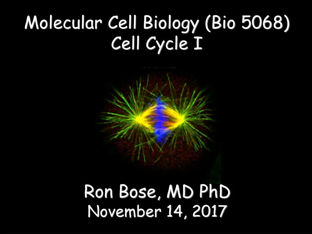
Molecular Cell Biology (Bio 5068) Cell Cycle I Ron Bose, MD PhD November 14, 2017
The cell cycle is primarily regulated by cyclically activated protein kinases Figure 17-15, 17-16 Molecular Biology of the Cell, 4th Edition
Overview of major cyclins and Cdks of vertebrates and yeast Table 17-1. Molecular Biology of the Cell, 4th Edition
Cdk activity is regulated by inhibitory phosphorylation and inhibitory proteins Why is cell cycle progression governed primarily by inhibitory regulation? Figure 17-18, 17-19. Molecular Biology of the Cell, 4th Edition
Cell cycle control depends on cyclical proteolysis Figure 17-20. Molecular Biology of the Cell, 4th Edition
Cyclin-dependent Kinase Inhibitor Proteins (CKI’s) • CIP/KIP family (p21Cip1, p27Kip1, p57Kip2): • Binds to Cdk2 and inhibits activity. • Binds Cdk4/6 and helps assemble complexes with cyclins. • INK4 family (p16, p15, p18, p19). • Specific for Cdk4 and Cdk6. • Binds Cdk subunit alone and prevents cyclin binding • Bind and inhibit Cdk4/6-Cyclin D heterodimers.
G1 Control M Cdk 4 & 6 Cyclin D1, 2, 3 INK4a proteins (p15,16, 18, 19) G2 Assembly & Sequestration G1 S Cdk2 Cyclin E Cip/Kip proteins (p21, p27, p57)
CHECKPOINTS IMPROPER SPINDLE ASSEMBLY M M DNA DAMAGE UNREPLICATED DNA STOP! G2 G2 G1 G1 S S DNA DAMAGE
Checkpoints: intracellular signaling pathways that determine if previous steps are complete before proceeding onto the next stage (complete DNA synthesis before entering mitosis; spindles must be assembled before exiting metaphase and entering into anaphase) and whether there has been any damage to the DNA. • DNA damage checkpoint: integrity of DNA • DNA damage is repaired before entering S, completing S or entering M. • DNA replication checkpoint: replication state of DNA • Complete DNA synthesis before mitosis. • Spindle assembly checkpoint: integrity of spindle • spindles must be assembled before exiting metaphase • into anaphase.
DNA DAMAGE RESPONSE PATHWAY M G2-PHASE CHECKPOINT STOP! G2 G1 S G1-PHASE CHECKPOINT S-PHASE CHECKPOINT
CELLULAR RESPONSES TO CHECKPOINT ACTIVATION (IR, etoposide, HU, gemcitibine, irinotecan, carboplatin…) CHECKPOINTS G1 S G2 M TEMPORARY CELL CYCLE ARREST & activation of DNA repair pathways APOPTOSIS SENESCENCE
DNA damage leads to cell cycle arrest in G1 Figure 17-33. Molecular Biology of the Cell, 4th Edition
Excessive stimulation of mitogenic pathways can lead to cell cycle arrest or cell death Figure 17-42. Molecular Biology of the Cell, 4th Edition
Molecular Biology of CancerMCB 5068 November 9, 2017Jason Weberjweber@DOM.wustl.edu
WHAT MAKES A CANCERCELL ACANCERCELL? UNLIMITEDGROWTH UNLIMITEDMOVEMENT
THE DEFINITION OF A TUMOR SUPPRESSOR Classical Features: Loss of function mutations Targeted allelic loss- Methylation or Deletion Inherited mutations that predispose to cancer Somatic mutation in spontaneous tumors Ability to inhibit transformed cells in vitro
Functional steps toward cancer Functional steps toward cancer Hanahan and Weinberg, Cell 2000
In the 1930’s while carrying out classic mutagenesis experiments in flies, Hermann Muller (Nobel, 1938) first noted that chromosome ends had “distinct” properties and named them telomeres (telo meaning ”end” and mere meaning “part”).
Telomere Function distinguishes between the chromosome end and a double strand break protects the chromosome from end-to-end fusions
100-400 nt single-strand overhang sub-telomeric telomeric double-stranded repeat region 10-20 kb Telomeres are found at the ends of chromosomes
Telomere Length 1 2 3 4 5 6 The “telomere” hypothesis (Harley and Greider) Senescence Population Doubling Time
Senescence Stop Rb p53 The telomere hypothesis Telomere Length Time
Other characteristics of senescent cells • Absence of proliferation markers • Flattened cell morphology • Increased cell volume • Senescence-associated β-galactosidase • p16INK4a tumor suppressor expression • Senescence-associated heterochromatin foci • Altered secretory profile (SASP)
Continued proliferation Crisis telomere dysfunction genetic catastrophe Rb p53 cell death What happens to telomeres if you bypass senescence??? Telomere Length Time
TP1 Dyskerin ? ? HSP90 Telomerase adds telomeric repeats to the 3’ termini of the chromosome -many components are required in vivo (this list continues to grow). hTERT p23 hTR CAAUCCCAAUC
ALT Stop hTERT Rb p53 Stable telomere maintenance The telomere hypothesis Senescence Telomere Length Crisis 1 in ~107 Time
The telomere hypothesis ALT Telomere Length Stop Crisis hTERT 1 in ~107 Time
T-ag t-ag SV40ER, TERT, and H-ras cooperate to transform normal human cells SV40 ER H-ras TERT Tumors Hahn et al, 1999
“Normal” Fibroblast Cancer -associated Fibroblast Pre-neoplastic Cell Tumorigenic Cell “Old/Senescent” Fibroblast “Normal” Fibroblast “Old” stroma is phenotypically similar to CAFs
Whether cells undergo telomere-based senescence or stress-induced senescence, p53 and Rb are the downstream effectors AND activation results in a permanent growth arrest (a.k.a potent tumor suppressor mechanism. Campisi 2005
Stem Cell Biology Jim Huettner 11/21/2017
Stem Cells: definition • Self Renewal - undifferentiated cells that can divide repeatedly while maintaining their undifferentiated state. • Pluripotency – ability to differentiate into a variety of different cell types
Types of Stem Cells Embryonic – from the inner cell mass of pre-implantation embryos, prior to formation of the 3 germ layers (ectoderm, mesoderm, endoderm) Somatic – undifferentiated cells found in specific locations in “mature” tissues iPS cells – induced pluripotent stem cells generated by reprogramming differentiated cells (or cell nuclei, i.e. therapeutic cloning)
Potency • Totipotent – able to generate every cell type including extraembryonic tissues • Pluripotent – able to generate cells from all three embryonic germ layers • Multipotent – able to generate a variety of cells from a particular somatic structure • Unipotent – only generate one cell type
Pluripotency markers • Stage-specific antigens: Anti-SSEA 3 and 4 recognize globo-series gangliosides • Tra1-60 and Tra1-81: keratin sulfate surface antigens • Oct3/4, Sox2, Nanog – transcription factors involved with maintaining pluripotency • Normal karyotype, and pre-X-inactivation?
Blastocyst chimera (+) High cloning efficiency Short doubling time Xa Xa Distal Oct4 enhancer High Nanog, Klf2/4, Rex1 Blastocyst chimera (-) Low cloning efficiency Long doubling time Xa Xi Proximal Oct4 enhancer Low Nanog, Klf2/4, Rex1 Two types of ES cells? “Naïve” (ICM-like) “Primed” (Epi-SC) Both types can self renew and give rise to cells from all 3 germ layers in teratomas or following in vitro differentiation
In vitro differentiation • Different culture conditions alter the fate of ES cells in vitro • Protocols exist for all three germ layers • Many, but not all, protocols involve aggregation of ES cells in “embryoid bodies” • Most protocols do not yield a single type of cell • Selection steps can help to remove undesired cell types • Need to ask: How far? & How faithful? • Pancreatic β cells: • Pagliuca FW, et al. (2014) Generation of Functional Human Pancreatic β Cells In Vitro. Cell. Oct, 159:428-39. • Rezania A, et al. (2014) Reversal of diabetes with insulin-producing cells derived in vitro from human pluripotent stem cells. Nat Biotechnol. Nov, 32:1121-33.
Reprogramming • SCNT – somatic cell nuclear transfer (reproductive and therapeutic cloning) – deterministic and fairly rapid • iPS – induced pluripotent stem cells – slow and stochastic (until recently) • Transdifferentiation – conversion of one terminally differentiated cell type into another without de-differentiation to an immature phenotype. Must rule out cell fusion or other explanations.
Cells of the Nervous System Bob Mercer rmercer@wustl.edu 362-6924
Neurons/Glia Neurons: electrically excitable cells that receive, process, and transmit information through electrical and chemical signals Glial Cells (neuroglia or glia): non-neuronal cells that maintain homeostasis, form myelin, and provide support and protection for neurons
Neurons Three basic neurons: Afferent Neurons (Sensory or Receptor Neurons): Respond to stimuli (touch, sound, light etc.) to send signals towards the spinal cord or brain. Efferent Neurons (Motor Neurons): Carry signals away from the central nervous system to generate a response (muscle contractions, glandular output). Interneurons: The largest group, relay signals from afferent to efferent neurons and to other interneurons within the same region of the brain or spinal cord in neural networks.
Types of Synapses • Electrical Synapse: • Allow low resistance pathways between cells through Gap Junctions • Found in cardiac muscle, smooth muscle • Allow fast conductive pathways • Chemical Synapse: • Physical gap between presynaptic cell membrane & postsynaptic cell membrane(Synaptic Cleft) • Chemical neurotransmitter released from presynaptic cell diffuses and binds to receptors on postsynaptic cell • Short delay (≈ ms) in conductive pathway
Neurotransmitters • Properties: • Transmit signals across a chemical synapse from one neuron to another "target" neuron, muscle cell, or gland cell • Synthesized by presynaptic cell • Released by presynaptic cell upon stimulation • If applied to postsynaptic membrane at physiological concentrations must elicit in vivo response • Many synthesized from simple and plentiful precursors such as amino acids and only require a small number of biosynthetic steps for conversion. • Exact numbers unknown, however more than 100 chemical messengers identified
Glial Cells • Functions: • Surround neurons and hold them in place • Supply nutrients, oxygen and other factors to neurons • To insulate one neuron from another • To destroy pathogens and remove dead neurons.