Download
1 / 23
400 likes | 1.86k Views
ESR = ERYTHROCYTE SEDIMENTATION RATE. Dr. MOHAMMED CHYAD AL-NOAEMI. What is the normal sedimentation rate?. The normal sedimentation rate (Westergren method): for males is 0-15 millimeters per hour, females is 0-20 millimeters per hour.

E N D
ESR = ERYTHROCYTE SEDIMENTATION RATE Dr. MOHAMMED CHYAD AL-NOAEMI
What is the normal sedimentation rate? The normal sedimentation rate (Westergren method): for males is 0-15 millimeters per hour, females is 0-20 millimeters per hour. The sedimentation rate can be slightly more elevated in the elderly. It is also increase in pregnancy and menstrual period.
The clinical usefulness of ESR Its increase indicates infection, or inflammation, or malignancy, or anemia. However, it rarely leads directly to a specific diagnosis.(i.e. a nonspecific test) It is considered as an index for assessment of severity of inflammatory conditions. ESR is increased according to the severity of the disease. It is used to monitor the response to therapy in certain inflammatory diseases. It is done if there is a suspicion of disease, the ESR may have some value as a "sickness index.“
Changes in ESR The ESR is increased by any cause or focus of inflammation (acute or chronic). However, it is useful in detecting and monitoring tuberculosis, certain forms of arthritis, autoimmune disorders, and diseases that cause vague symptoms. Diseases in general are either mild, moderate or sever. Therefore, ESR is increased according to the severity of the disease. The ESR is decreased in sickle cell anemia, polycythemia, and congestive heart failure.
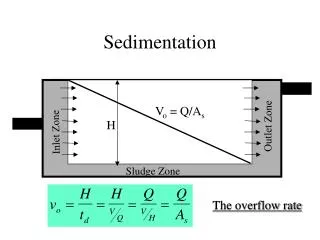
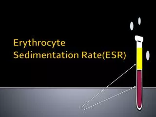
The erythrocyte sedimentation rate (ESR) Is also called a sedimentation rate, sed rate, is the rate at which red blood cells precipitate in a period of 1 hour. It is a common haematology test which is a non-specificmeasure of inflammation. To perform the test, anticoagulatedblood is placed in an upright tube, known as a Westergren tube and the rate at which the RBCfall is measured and reported in mm/h.
Method (Westergren Tube) Blood is obtained by clean venepuncture, and thoroughly mixed in a proportion of 4 volumes of blood (as 2ml) to 1 volume of filtered solution of sodium citrate (0.5ml). Or 2ml of EDTA anti-coagulated blood + 0.5ml of diluting fluid as normal saline (0.9%NaCl). Blood is mixed thoroughly with the anticoagulant by gentle repeated inversion. A clean dry Westergren tube is filled and adjusted to the 0 mark. The tube is then placed in a strictly vertical position under room temperature (18-25C). After one hour the distance from zero to the top of the column of sedimenting red cells is read in mm and recorded as ESR value. The result is expressed as: ESR= ? mm/hour.
Wintrobe Method This picture shows a rack holding Wintrobe tubes, in which anticoagulated whole blood has just been added. (Time: 0)
Wintrobe Method Red blood cells have settled, leaving plasma at the top of the tube. Reading: 18 mm/hour (Time: one hour)
Method (WintrobeTube) By putting Anticoagulated blood directly into the wintrobe tube (without dilution). It is used for demonstration. The normal values will be slightly less than westergren method. [This method is less accurate than the westergrenmethod.Br Med J. 1951 December 22; 2(4746): 1496–1497.Westergren and Wintrobe Methods of Estimating E.S.R. Compared.David Gilmour and A. J. Sykes].
Wintrobe method: The Wintrobe method is performed similarly except that the Wintrobe tube is smaller in diameter than the Westergren tube and only 100 mm long. EDTA anticoagulated blood without extra diluent is drawn into the tube, and the rate of fall of red blood cells is measured in millimeters after 1 hour. The shorter column makes this method less sensitive than the Westergren method because the maximal possible abnormal value is lower. However, this method is more practical for demonstration purposes. http://www.medicine.mcgill.ca/physio/vlab/bloodlab/esr.htm
Mechanism of ESR Formation The ESR is governed by the balance between pro-sedimentation factors, mainly fibrinogen, and those factors resisting sedimentation, namely the negative charge of the erythrocytes. When an inflammatory process is present, the high proportion of fibrinogen in the blood causes red blood cells to stick to each other. The red cells form stacks called 'rouleaux' which settle faster. Rouleaux formation can also occur in association with some lymphoproliferative disorders in which one or more immunoglobulins are secreted in high amounts.
http://www.medicine.mcgill.ca/physio/vlab/bloodlab/esr.htm Erythrocytes aggregate in a special way, forming rouleaux. Rouleaux are stacks of erythrocytes which form because of the unique discoid shape of the cells. The flat surface of the discoid RBCs give them a large surface area to make contact and stick to each other; thus, forming a rouleaux. Normal distributed RBC Precipitated RBC by rouleaux formation in ESR tube
Rouleaux Formation The RBC's here have stacked together in long chains. This is known as "rouleaux formation" and it happens with increased serum proteins, particularly fibrinogen and globulins. Such long chains of RBC's sediment more readily. This is the mechanism for the sedimentation rate, which increases non-specifically with inflammation and increased "acute phase" serum proteins.
ESR (mm/hr) reference ranges from a large 1996 study Age (years) 205590 Men 10 15 19 Women 15 21 23 Normal values of ESR have been quoted as 1 to 2 mm/hr at birth, rising to 4 mm/hr 8 days after delivery, and then to 17 mm/hr by day 14.
Normal Values(mm/hr. = millimeters per hour) The widely used rule for calculating normal maximum ESR values in adults (98% confidence limit) is given by a formula devised in 1983
Materials Anticoagulant-Diluent Solution Trisodium citrate dehydrate (31.3 gram/L -DW) The solution is filtered through a sterile membrane, maximum pore diameter 0.22 mm, into a sterile container. It must be discarded if turbid. Westergren tube is graduated from 0-200mm. Blood sample.
Is there anything else we should know? • ESR and C-reactive protein (CRP) are both markers of inflammation. Generally, ESR does not change as rapidly as does CRP, either at the start of inflammation or as it goes away. CRP is not affected by as many other factors as is ESR, making it a better marker of inflammation. However, because ESR is an easily performed test, many doctors still use ESR as an initial test when they think a patient has inflammation. • If the ESR is elevated, it is typically a result of globulins or fibrinogens. Your doctor may then order a fibrinogen level (a clotting protein that is another marker of inflammation) and a serum protein electrophoresis to determine which of these (or both) is causing the elevated ESR.
Relation to C-reactive protein C-reactive protein is an acute phase protein that is produced by the liver during an inflammatory reaction. Since C-reactive protein levels in the blood rise more quickly after the inflammatory or infective process begins, ESR is often replaced with C-reactive protein measurement. There are specific drawbacks, however, as they were found to be independently associated with a diagnosis of acute maxillary sinusitis so that the combination of the two measurements improved diagnostic sensitivity and specificity.
REFERENCES 1. Practical Physiology. Mohammed Chyad Al-noaemi 2. J ClinPathol. 1973 April; 26(4): 301–302. • PMCID: PMC477708 3. The most satisfactory method of performing the test was introduced by Westergren in 1921. [Saadeh C. The erythrocyte sedimentation rate: old and new clinical applications. South Med J. 1998;3:220–5.]. 4.http://www.ncbi.nlm.nih.gov/pmc/articles/PMC477708/?page=1