Download
1 / 18
180 likes | 312 Views
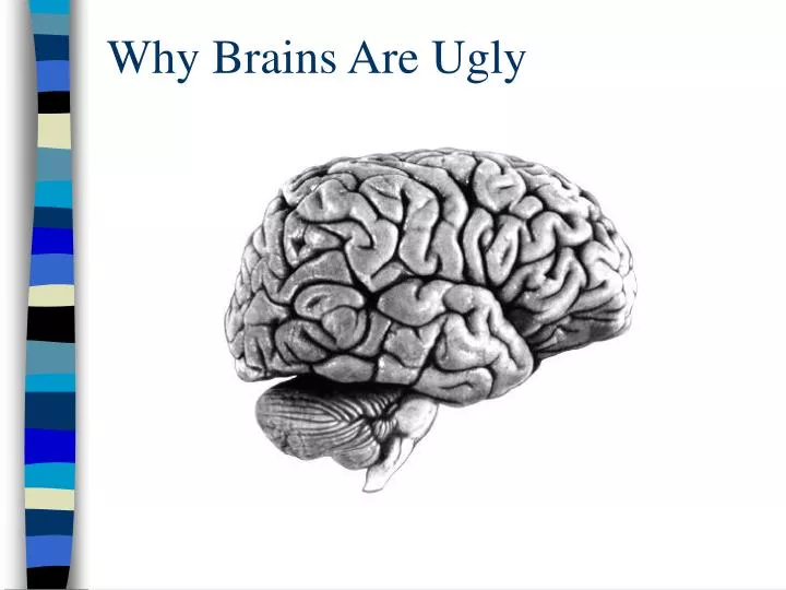
Why Brains Are Ugly. brains are ugly and complicated looking. Let’s face it. And we may be the worst offender!. F. P. F. O. T. The problem is that much of the ‘architecture’ of the brain is not functionally meaningful. Santiago Ramon y Cajal.

E N D
brains are ugly and complicated looking Let’s face it. . .
And we may be the worst offender! F P F O T The problem is that much of the ‘architecture’ of the brain is not functionally meaningful
Santiago Ramon y Cajal ‘…to extend our understanding of neural function to the most complex human physiological and psychological activities, it is essential that we first generate a clear and accurate view of the structure of the relevant centers, and of the human brain itself, so that the basic plan – the overview – can be grasped in the blink of an eye.’ Histology of the Nervous System of Man and Vertebrates (1909)
Telencephalon (or ‘Cerebrum’) Brainstem To Begin, Let’s Break the Brain into Two Functional Parts. Swanson, 1995
Telencephalon Now add in some sub-divisions Cortex Limbic System Basal Ganglia Diencephalon Thalamus Hypothalamus Midbrain Superior Colliculus Inferior Colliculus Brainstem Cerebellum Brain Regions You Should KNOW Hindbrain Spinal Cord
81 Tel: 38 Di: 6 Mid: 4 Hind: 7 Cblm: 10 SC: 35 Rat CNS Human CNS Swanson, 1995 Tel: 81 Di: 4 Mid: 1 Hind: 2 Cblm: 10 SC: 2
64 Tel: 64 Di: 6 Mid: 10 Hind: 6 Cblm: 10 SC: 4 Tel: 38 Di: 6 Mid: 12 Hind: 7 Cblm: 11 SC: 26 Zebra Finch CNS Pigeon CNS
Rat Pigeon Finch Human Telencephalon: 38 38 64 81 Diencephalon: 6 6 6 4 Midbrain: 4 12 10 1 Hindbrain: 7 7 6 2 Cerebellum: 10 11 10 10 Spinal Cord: 35 26 4 2
S e n s o r y – D o r s a l Thalamus M o t o r – V e n t r a l Hypothalamus The Brainstem has a simple dorsal/ventral organization Dorsal When they are sick, Some Dogs Might Vomit Ventral Note the two sub-divisions of the diencephalon: Thalamus and Hypothalamus
Telencephalon Stimulus (what), motor control Primary Motor Cortex Primary Sensory Cortex Rat CNS Motor output Sensory input Thalamus relay, feedback (except olfaction) Midbrain Stimulus (where) vision and audition only andorienting responses Spinal Cord reflexes, input, output GENERAL Overview of Sensory System Organization (L/R and R/L crossing for vision, touch, audition only)
Rat CNS Telencephalon Primary Auditory Cortex Thalamus Medial Geniculate Midbrain Inferior Colliculus Hindbrain Pons, Medulla
Telencephalon TELENCEPHALON AND DIENCEPHALON (some key subregions for S&P) Cortex Limbic Basal Ganglia Cortex Diencephalon Hippocampus Amygdala Thalamus Basal Ganglia Hypothalamus Thalamus Hypothalamus
1 2 3 From Thalamus 4 Back ToThalamus 5 6 Output Layer CORTEX is made of Vertical Columns, each containing 6 layers of neurons
Primary Cortex Thalamus 2D Receptor Array However…
Cortex Is ‘Modular’ Means the size and density of cortical columns is fixed Thalamus Dense 2D Receptor Array
Because Cortex is Modular, Sensory ‘Maps’ in Primary Cortex are Distorted by Variation in the Density of Sensory Receptors
Ha-ha! That looks funny! A ‘homunculus’ of the USA