Download
1 / 15
170 likes | 527 Views
Lecture 5. Integumentary System. Integumentary System. Consists of: Skin (Epidermis & Dermis) Accessory structures Hair Nails Glands Functions Protection Sensation Temperature regulation Vitamin D production Excretion. Fig. 5.1. Epidermis and Dermis. Fig. 5.2. Epidermal Cells.

E N D
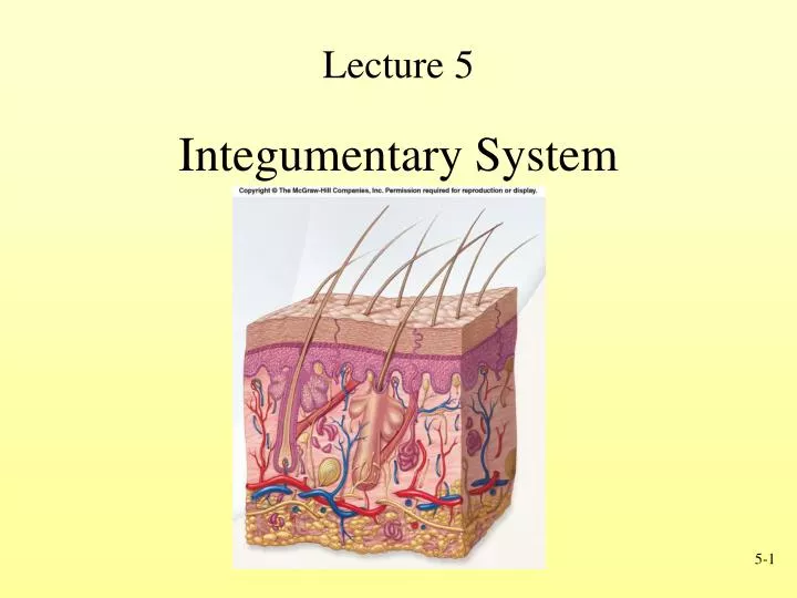
Lecture 5 Integumentary System
Integumentary System • Consists of: • Skin (Epidermis & Dermis) • Accessory structures • Hair • Nails • Glands • Functions • Protection • Sensation • Temperature regulation • Vitamin D production • Excretion Fig. 5.1
Epidermis and Dermis Fig. 5.2
Epidermal Cells • Cell types • Keratinocytes: Produce keratin for strength • Melanocytes: Contribute to skin color (pigment melanin) • Langerhans’ cells: Part of the immune system • Merkel’s cells: Detect light touch and pressure Fig. 5.2 • Keratinization: Cells die and produce outer layer that resists abrasion and forms permeability layer
Epidermal Strata • Stratum Basale • Deepest portion of epidermis and single layer • High mitotic activity • Stratum Spinosum • Limited cell division • Stratum Granulosum • In superficial layers nucleus and other organelles degenerate and cell dies Fig. 5.2
Epidermal Strata • Stratum Lucidum • Thin, clear zone • Stratum Corneum • Most superficial and consists of cornified cells • Squamous in shape, filled with keratin Fig. 5.2
Dermis • Structural strength • Two layers • Deeper layer - dense connective tissue; stretch marks (striae) • Superficial layer underneath epidermis - loose connective tissue Fig. 5.6
Review Question The layer of keratinocyte cells of the epidermis with the highest rate of cell division is the • Stratum spinosum • Stratum basale • Stratum corneum • Stratum granulosum • Stratum lucidum
Hypodermis • Skin rests on this, but not a part • Also called • Subcutaneous tissue • Superficial fascia • Consists of loose connective tissue • Types of cells • Fibroblasts • Adipose (fat) cells • Macrophages • Subcutaneous fat Fig. 5.6
Accessory Skin Structures • Hair • Found everywhere on human body except palms, soles, lips, nipples, parts of external genitalia, and distal segments of fingers and toes • Nails • Glands • Sebaceous or oil glands • Sudoriferous or sweat glands
Hair Structure • Composed of shaft and root • Shaft protrudes above skin surface • Root located below surface and base forms the hair bulb • Hair follicle consists of layer of dermis and epidermis • Arrector pili • Smooth muscle • Raises hair Fig. 5.9
Nails • Anatomy • Nail root proximally • Nail body distally: Eponychium or cuticle • Growth • Grow continuously unlike hair Fig. 5.8
Oil and Sweat Glands • Sebaceous glands • Produce sebum • Oils hair and skin surface • Sudoriferous glands (sweat) • Most commonly found in palms, soles and forehead • Also found in axillae (arm pit), genitalia, around anus • Ceruminous glands (cerumen or ear wax) • Mammary glands Fig. 5.10
Points to Remember • Two components to integumentary system • Skin (epidermis & dermis) • Accessory structures (hair, nails and glands) • Keratinization (accumulation of keratin) produces upper layer of cells of epidermis, hair and nails • Two types of glands in skin • Sebaceous (oily) • Sudiferous (watery)


![[lecture#5]](https://cdn0.slideserve.com/109460/slide1-dt.jpg)