Download
1 / 15
150 likes | 304 Views
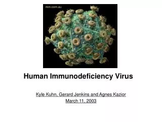
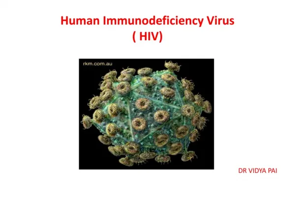
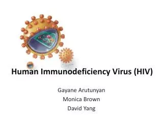
HUMAN IMMUNODEFICENCY VIRUS (HIV). INTRODUCTION. HIV VIRUS IS HUMAN IMMUNODEFICENCY VIRUS IT CAUSES (ACQUIRED IMMUNODEFICENCY SYNDROME)(AIDS)

E N D
INTRODUCTION • HIV VIRUS IS HUMAN IMMUNODEFICENCY VIRUS IT CAUSES (ACQUIRED IMMUNODEFICENCY SYNDROME)(AIDS) • HIV VIRUS exists as roughly spherical particles surface pf each particle has studded spikes outside the human body called as virions or in infected immune cells
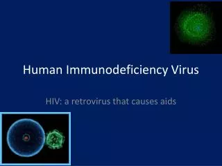
TAXONMIC STATUS: • GROUP: GROUPVI (ss RNA_RT) • FAMILY: RETROVIRIDAE • GENUS: LENTIVIRUS • The viruses of Lentivirus family cause diseases in monkeys cattle sheep, cats and goats. HIV is a retrovirus .it is host specific virus. It was discovered in monkeys for the first time but it does not affect the monkeys rather causes aids in humans
It has two types HIV- 1 AND HIV- 2. TYPE 1 is highly infectious and causes 90%of the infection globally. HIV-2 is found in west Africa and confined there TYPES OF HIV VIRUS:
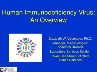
MORPHOLOGY • SIZE: • 0.1micron • 1/20th of the length of an inch • It is too small to be seen with an a compound microscope and it is visible with the electron microscope.
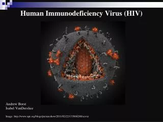
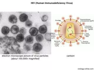
Structure: HIV is a retrovirus It has an enzyme (reverse trancriptase which converts single stranded RNA to double stranded DNA) It has a coat of fatty material called as viral envelope or capsid It has 72 little spikELTS projecting from the capsid.
ENZYME AND PROTIEN STRUCTURE • VIRAL ENVELOPE: • The envelope is made up of gp120 and gp 4 and gp4 .just below the viral layer is made from protein p17. • Capsid is made up of p24 it is bullet shape. • Enzymes • Integrase • Protease • Reverse transcriptase
Genome The HIV virus is a retrovirus it has single stranded RNA in its genome and it does not have a complex genome it ois comprised of 9 genes only • 3 are (gag pol env) these are encoded to make new virus particles • The rest 6 (tat ,rev ,nef, vif, vpr, and vpu) are used in the formation of proteins that control the ability of HIV to infect a cell and cause disease
Lifecycle of HIV virus: • Entry: The first step of the lifecycle of HIV is its attachment on the CD4 receptor site of the helper Tcell where it s spikes stick to the protein of CD4 site • REVERSETRANSCRIPTION AND INTEGRATION: once inside the cell the HIV enzyme REVERSETRANSCRIPTASE converts the single stranded mRNA into double stranded DNA using the host genome which is compatible to host genome .The enzyme INTEGRASE slices the human dna into viral DNA.
Transcription and translation: HIV is a provirus it stays in host genome for a long time .It becomes activated and expressed in the same way as the human genes .the mRNA is used as blue print to make new HIV virus copies viral proteins and enzymes . Assembly budding and integration: when the cell produces and translates mRNA copies they are the complete copies of HIV material and they gather together with the newly made HIV proteins and enzymes to form new viral proteins which are released from the cell .Enzyme Protease plays an important role in chopping up of DNA strands to make anew viral core. the new vcirions are ready to infect other cells and they grow and mature by body fluids.
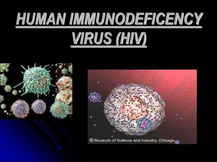
Electron microscope Electron Microscope slide showing virus transmission from cell membrane of T helper cell