Download
1 / 1

E N D
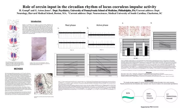
Introduction Activation of noradrenergic locus coeruleus (LC) neurons promotes wakefulness and behavioral arousal. In rats, the LC receives circadian inputs via a relay circuit from the suprachiasmatic nucleus (SCN) through the dorsomedial hypothalamus (DMH); this circuit input increases LC activity during the active period (Aston-Jones et al., Nat Neurosci 2001, Aston-Jones et al., 2005). Recent evidence from out lab indicates that this circuit mediates in part the circadian regulation of the wake-sleep cycle (Gonzalez and Aston-Jones, 2006). DMH neurons expressing the peptide neurotransmitter orexin / hypocretin are ideally situated to act as a relay between the SCN and the LC due to their synaptic inputs from the SCN and innervation of the LC. Here we examine the hypothesis that orexin is involved in transmitting circadian signals to the LC using single-unit recordings of LC neurons in anesthetized rats maintained in 12:12 light-dark housing. METHODS Adult male Sprague Dawley rats were kept on a 12 hr LD cycle for at least one week. Under halothane anesthesia administered by a tracheal cannula, animals were placed in stereotaxic frame with incisor bar lowered to place skull at approximately 12 ° from the horizontal plane. “Double barrel” micropipettes were manufactured by fusing an infusion micropipette (12-20µm outer diameter) to a recording micropipette at its shank so that its tip was approximately 100 µM recessed from the recording pipette tip. The recording micropipette (tip diameter, 2-4 µm; 10-20 MΩ impedance) was filled with 2 % Pontamine Sky Blue dye in 0.5 M Na-acetate, while the infusion pipette was filled with drug dissolved in aCSF. A 5-mm-diameter hole was drilled in the skull above the LC, the dura reflected and the “double-barrel” pipette was aimed 4 mm caudal to lambda, 1.2 mm lateral to midline, and approximately 5.8-6.5 mm ventral to the skull surface. LC neurons were tentatively identified using well established criteria: an entirely positive, > 2 ms notched waveform in unfiltered recordings, and slow tonic discharge (0.5-6 Hz) except following noxious stimuli (phasic activation for 0.5-1 s). Microinjections were made by pneumatic pressure. After isolating a single LC neuron, baseline firing frequencies were recorded for at least 150 sec. Solutions were administered by applying a single pulse of 30-100 msec duration at 40 psi every 15 seconds so that each pulse was approximately 12 nl and the total volume applied was 120-180 nl over a period of 150 - 300 sec. At the end of recordings, micropipette penetrations were marked by iontophoretic ejection of dye with negative current pulses (20 µA, 50 % duty cycle for 15 min.), brains snap-frozen in isopentane at -80 °C and coronal sections through the LC (40 µm thick) were cut on a cryostat, mounted and stained with neutral red. SUMMARY The results strongly suggestLC neurons receive circadian timing information from orexin neurons. Results presented here and elsewhere also implicate orexin neurons located in the dorsomedial hypothalamus Role of orexin input in the circadian rhythm of locus coeruleus impulse activity H. Gompf1 and G. Aston-Jones2. Dept. Psychiatry, University of Pennsylvania School of Medicine, Philadelphia, PA;1Current address: Dept. Neurology, Harvard Medical School, Boston, MA; 2Current address: Dept. Neurosciences, Medical University of South Carolina, Charleston, SC B. A. Active phase Rest phase * * Figure 2: Is SB338467-mediated inhibition of LC firing rate associated with increased activation of dorsomedial orexin neurons? A: FOS expression in orexin-positive neurons was counted in animals sacrificed 90 minutes after ZT 9-10, 11-12, or 14-15 in the dorsomedial hypothalamus (top panel). Average counts in the dorsomedial hypothalamus (DMH) are shown in white bars and lateral hypothalamus (LH) average counts are shown in dark bars (lower panel). FOS immunoreactivity in orexin-positive neurons was significantly greater in the DMH at ZT 11 – 12 (49.4 ± 2.25 %; n = 5) than at ZT 9 – 10 (20.9 ± 2.5%; n = 6; p < 0.001) and not different from orexin neurons at ZT 14-15 (49.45 ± 3.13 %; n = 4). This difference is not seen in orexin neurons in the LH (27.2 ± 3.5 % vs 26.3 ± 1.9 %, respectively; p = 0.816). An increase in FOS immunoreactivity in orexin neurons was observed in both the DMH (49.5 ± 3 %, n = 6, p < 0.001) and LH (34.3 ± 2.6 %; p = 0.1) later in the active phase (ZT 14 – 15), though the increase in the LH is not significant. B: To examine whether increased activation of medial orexin neurons translates into increased activation of LC impulse activity, we recorded LC neurons between ZT 11 – 12. Average firing frequency (2.9 ± 0.4 Hz, n = 16) during this time period was higher than firing frequencies during the rest of the light period (p = 0.04). Similar to neurons recorded during the active phase, SB-338467-A inhibition of fast-firing neurons was observed during the transition period from ZT 11 – 12 (fast: 3.6 ± 0.4 Hz control, 2.0 ± 0.5 Hz SB-338467-A, n = 10, p = 0.02; slow: 1.7 ± 0.2 Hz control, 1.2 ± 0.4 Hz SB-338467-A, n = 6, p = 0.2, figure 4), indicating that immediately prior to the onset of the active phase a subset of LC neurons behave similarly to neurons recorded in the middle of the active phase. References: Aston-Jones, G., Chen, S., Zhu, Y., Oshinsky, M.L. A neural circuit for circadian regulation of arousal. Nat. Neurosci. 4:732-8 (2001). Aston-Jones, G., Card, J.P., Zhu, Y., Gonzalez, M. and Haggerty, E. The NE system as a target for hypocretin neurons: Implications for regulation of arousal. In: Hypocretins: Integrators of Physiological Functions, L. DeLecea and G. Sutcliffe, eds. Kluwer / Springer (2005), pp 137-152. Gonzalez, M. and Aston-Jones, G. Circadian regulation of arousal: Role of the noradrenergic locus coeruleus system and light exposure. Sleep 29:1314-23 (2006). Sites of DMH lesions that led Aston-Jones et al (2001) to propose a SCN-DMH-LC relay circuit responsible for transmission of circadian temporal information to the LC. These excitotoxic lesions eliminated circadian rhythms in LC impulse activity Aston-Jones et al (2001). Location of orexin neurons in the hypothalamus. Neurons expressing orexin can be found in the lateral (LH), perfornical and dorsomedial hypothalamus (DMH). Figure 1: LC neuronal impulse activity during the rest (A, C) and active phase (B, D). Average impulse activity was greater during the active period than during the rest period in LC neurons (ZT 5 - 10: 1.8 ± 0.2 Hz, n = 30; ZT 14-20: 2.9 ± 0.4 Hz, n = 19, p < 0.05). Local microinfusion of ACSF had no effect on LC firing frequency (1.9 ± 0.6 Hz control vs 1.8 ± 0.5 Hz, n = 6, p = 0.36 ACSF). However, microinfusion of 100 µM orexin-A significantly increased LC firing frequency in all neurons recorded (1.5 ± 0.5 Hz control vs 3.5 ± 0.9 Hz 100 µM orexin A, n = 6, p = 0.009). Raw traces (A, B) and composite firing frequency responses (C, D) of LC neurons to the orexin A antagonist SB338467 are shown. Whereas no significant change in firing frequency was observed in average LC firing frequency during SB-338467-A infusion during the rest period (1.9 ± 0.3 control, 2.2 ± 0.4 SB-338467-A, n = 17, p = 0.4), SB-338467-A infusion during the active period significantly decreased LC firing frequencies (2.9 ± 0.4 Hz control, 1.8 ± 0.3 Hz SB-334867-A, n = 19, p = 0.01). Median firing frequencies during the rest period were 2.3 Hz (C). During the active period (D), neurons with firing frequencies faster than this were more likely to be inhibited by SB338467 than neurons firing slower (fast: 4.2 ± 0.6 Hz control, 1.6 ± 0.5 Hz SB-334867, n = 9, p < 0.0001; slow: 1.7 ± 0. 1Hz control, 1.9 ± 0.3 Hz SB-334867, n = 10, p = 0.8). This difference was not observed during the rest period (fast: 3.7 ± 0.3 Hz control, 4.0 ± 0.5 Hz SB-334867, n = 5, p = 0.3; slow: 1.2 ± 0.2 Hz control, 1.4 ± 0.3 Hz SB-338467-A, n = 12, p = 0.3). SCN DMH Orexin neurons LC Wakefulness, Arousal Identification of recording site by Pontine Sky Blue and Nissl staining. Supported by PHS NS24698