Download
1 / 23
230 likes | 431 Views
X-ray PhotoEmission Electron Microscopy of domains in GaMnAs. I06 Diamond Light Source Stuart Cavill Sarnjeet Dhesi Francesco Maccherozzi Kevin Edmonds Robin Marshall James Haigh. X-ray PhotoEmission Electron Microscopy. In normal XMCD measurements, the difference in the x-ray

E N D
X-ray PhotoEmission Electron Microscopy of domains in GaMnAs I06 Diamond Light Source Stuart Cavill SarnjeetDhesi Francesco Maccherozzi Kevin Edmonds Robin Marshall James Haigh
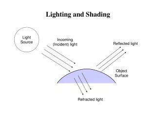
X-ray PhotoEmission Electron Microscopy • In normal XMCD measurements, the difference in the x-ray • absorption spectra for positive and negatively circular polarised • x-rays is measured, and difference in absorption is proportional • to the magnetic moment in the direction of the polarisation. • The absorption is measured by secondary process: • Auger emission of electrons, by measuring the drain current. • Fluorescence, by measuring the intensity of light emitted • by the sample under illumination with x-rays. • In PEEM, we measure the emitted electrons • from the sample by accelerating them away • from the sample and focusing the image onto • a fluorescent screen. • This gives a measure of the local x-ray absorption. • Can measure in positive and negatively • circular polarised light to get local XMCD. camera fluorescent screen 0V e- optics, analyser e- energy hν -2kV
X-ray PhotoEmission Electron Microscopy L3 3. 2. off peak, negative circular 1. on peak, negative circular 1. L2 2. 4. 4. off peak, positive circular 3. on peak, positive circular energy
ave 22948-22972 • 20mm FOV • = 0 (107 on manip) T=-175C
10mm FOV • = 5 (102 on manip) T=-172C • 20mm FOV • = 5 (102 on manip) T=-172C ave 22992-22307 ave 23010-23025
20mm FOV • = 5 (102 on manip) T=-172C ave 23128-23143
OOMMF simulation of domains. • Initial state is with each element having a random direction of magnetisation. • State is allowed to evolve to minimise energy. • Domains form as different regions have different direction magnetisation. • The domains grow and merge.
LEEM image. 25nm Ga(1-x)MnxAs , x = 0.12
a asymmetry (%) c b a b d asymmetry (%) d c
Heating above Tc 1 2 3 4 5 1 2 3 4 5
below Tc below Tc above Tc Cool Heat