Download
1 / 21
260 likes | 722 Views
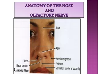
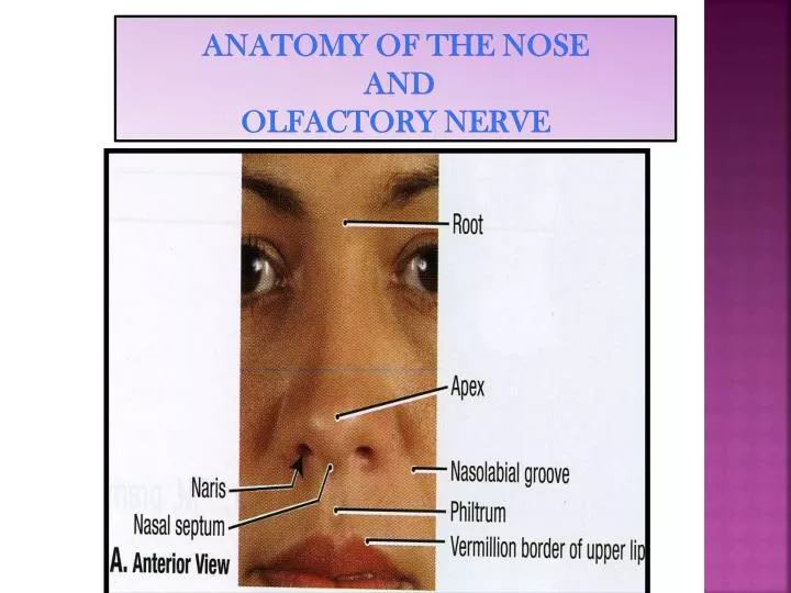
ANATOMY OF THE NOSE AND OLFACTORY NERVE. OBJECTIVES. By the end of this lecture the students should be able to: Describe the structures forming the walls of the nasal cavity. List the main structures draining into the lateral wall of the nasal cavity.

E N D
OBJECTIVES • By the end of this lecture the students should be able to: • Describe the structures forming the walls of the nasal cavity. • List the main structures draining into the lateral wall of the nasal cavity. • Differentiate between the respiratory and olfactory regions of the nasal cavity. • List the main sensory and blood supply of the nose. • Describe the olfactory pathway.
Nasal cavity • It extends from nostrils anteriorly to the choanae posteriorly. • Divided into right and left parts by the nasal septum. • Each part has: • Roof • Floor • Lateral and • Medial walls.
Floor • Formed by: • Nasal (upper)surface of the hard (bony) palate: • Palatine process of maxilla, anteriorly. • Horizontal plate of the palatine bone, posteriorly.
Roof • Formed by: • Body of sphenoid, posteriorly. • Cribriform plate of ethmoid, in the middle. • Frontal, and nasal bones. Anteriorly.
Medial wall • The nasal septum : • Vertical plate of ethmoid. • Septal cartilage. • Vomer.
Lateral wall • Marked by: • 3projections; (Nasal Conchae). • Superior, middle, and inferior Nasal Conchae • The space below each concha is called Meatus. • Superior, middle, and inferior meatus. • The space (fossa) above the superior concha is the Sphenoethmoidal recess.
PARANASAL SINUSES • They are cavitiesinside the: • Frontal bone • Sphenoid bone • Ethmoid bone • Maxilla • They are: • Lined with mucoperiosteum; • Filledwith air; • Communicate with the nasal cavity. • Open in the lateral wall of the nasal cavity • Function: • Lighten he skull weight • Amplify the sound as we speak.
SINUSES opening in lateral wall • Sphenoethmoidal recessreceives the opening of sphenoidal air sinus • Superior meatus;receives the opening of posterior ethmoidal sinus. • Middle meatus; contains bulla ethmoidalisand hiatus semilunaris, • Receives the openings of maxillary,frontal, & anterior, middle ethmoidal sinuses. • Inferior meatus; receives the opening of nasolacrimalduct.
The mucosal lining of these sinuses is continuous with that in the nose and the throat. So infection in these area tends to migrate into the sinuses causing sinusitis. • Note : all sinuses open into the middle meatusEXCEPT: • Sphenoidal sinus : in sphenoethmoidal recess. • Posterior ethmoidal sinus : in superior meatus.
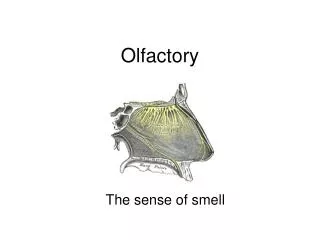
Nasal mucosa • Olfactory : • It is delicate and contains olfactory nerve cells. • It is present in the roof,lateral wall and upper part of nasal cavity. • On the lateral wall, it lines the upper surface of the superior conchaand the sphenoethmoidal recess. • On the medial wall, it lines the superior part of the nasal septum.
RESPIRATORY MUCOSA • It is thick, ciliated highly vascular and contains mucous glands & goblet cells • It lines the lower part of the nasal cavity (from skin of vestibule to the superior concha). • It functions to moisten, clean and warm the inspired air. • The air is moistenedby the secretion of numerous serous glands. • It is cleanedby the removal of the dust particles by the ciliary action of the columnar ciliated epithelium that covers the mucosa. • The air is warmedby a submucous venous plexus.
Nerve supply • The nerves of General Sensation are derived from the Ophthalmic & Maxillarydivisions of trigeminal nerve. • The anterior part is supplied by:Anterior Ethmoidal nerve. • The posterior part is supplied by: • 1-Nasopalatine, • 2- Nasal, and • 3- Palatine branches of the pterygopalatine ganglion.
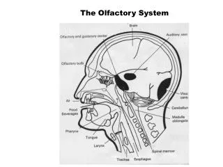
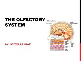
Olfactory pathway • 1st neurone: • Olfactory receptors are specialized, ciliatednerve cells that lie in the olfactory epithelium. • The axons of these bipolar cells 12-20 fibers form the true olfactory nerves fibers. • Which passethrough the cribriform plateof ethmoid. • They join the olfactory bulb
Preliminary processingof olfactory information is within the olfactory bulb, which contains interneurones and large Mitral cells;axons from the latter leave the bulb in the olfactory tract.
Olfactory pathway • 2nd neurone: • It is formed by the Mitral cells of olfactory bulb. • The axons of these cells form the olfactory tract. • Each tract divides into 2 roots at the anterior perforated substance: • Lateral root: • Carries olfactory fibers to end in cortex of the Uncus & adjacent part of Hippocampalgyrus(center of smell).
Medial root : • crosses midline through anterior commissure and joins the uncrossed lateral root of opposite side. • It connects olfactory centers of 2 cerebral hemispheres. • So each olfactory centre receives smell sensation from both halves of nasal cavity. • NB. Olfactory pathway is the only sensory pathway which reaches the cerebral cortex without passing through the Thalamus.
Arterial supply • Sphenopalatine artery (maxillary) . • Ethmoidal anterior and posterior (ophthalmic). • Superior labial (facial). • Applied anatomy : • Rich arterial anastomosis on anterior & inferior part of nasal septum (Little’s area) is the most common site for epistaxis.
Venous drainage • Plexus in sub mucosa by veins accompanying the arteries
Lymph drainage • Submandibular nodes • Upper deep cervical nodes.