Download
1 / 28
290 likes | 299 Views
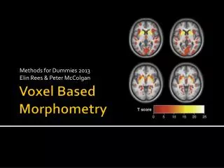
Voxel-based Morphometric Analysis. Martinos Center for Biomedical Imaging, Massachusetts General Hospital, Charlestown, MA, United States. Outline. Voxel-based Morphometry Surface-based Analysis Comparisons. Voxel-based Morphometry. Anatomical Changes. GM. Subject 1 88 yo.

E N D
Voxel-based Morphometric Analysis Martinos Center for Biomedical Imaging, Massachusetts General Hospital, Charlestown, MA, United States
Outline • Voxel-based Morphometry • Surface-based Analysis • Comparisons
Anatomical Changes GM Subject 1 88 yo Subject 2 19 yo
Voxel-based Morphometry (VBM) How do the sizes of gray/white matter and CSF structures change between subjects/populations? GM WM CSF Subject 1 88 yo Subject 2 19 yo
Voxel-based Morphometry (VBM) How to define a volume without defining a boundary? How to compare regions without defining a region? WM CSF
Non-linear Spatial Normalization Subject 2 (Target) Subject 1 Subject 1 Subject 2 CSF • Eyes move closer together • Lips curl and get wider
Non-linear Spatial Normalization Subject 2 (Target) Subject 1 Subject 1 Subject 2 CSF • Eyes move closer together • Lips curl and get wider
Keep Track of Changes in Size CSF • Voxel on the left side of mouth does not change size • Voxel on the right gets much larger • Eyes do not change size • Quantification: Gray Matter “Density”
Jacobian Jacobian Subject 2 (Target) Subject 2 (Target) Subject 1 CSF • Map of change in volume at each voxel in target space • Eyes are 0 • Left side of lips are 0 (black) • Right side of lips are yellow (expansion)
GM WM CSF Normalization and Segmentation Individual T1 (Template Space) Jacobian Individual T1 Spatial Normalization Expansion Compression Group Template (Target) Segmentations Values between 0 and 1 “Density”, Partial Volume Note: in FSL, Segmentations computed in native space “Unified Segmentation”, Ashburner and Friston, NI, 2005 “Optimized VBM” Good, et al, NI, 2001. Douaud, et al, Brain. 2007.
Modulation and Smoothing (Template Space) GM Segmentation (Concentration) Multiply 3D Smooth GM Density Jacobian
Aging Gray Matter Volume Study Positive Age Correlation Negative Age Correlation N=40 Statistical Maps (SPM8/VBM8) p<.01 GM Density
Surface-based Analysis: Cortex • Outer layer of gray matter • White/Gray Surface • Pial Surface
Cortical Thickness pial surface • Distance between white and pial surfaces along normal vector. • 1-5mm
IndividualThickness Maps 46 yo 88 yo 18 yo Male Female Salat, et al, 2004, Cerebral Cortex
A Surface-Based Coordinate System Common space for group analysis (like Talairach). Fischl, et al, 1998, NI.
Surface Spatial Smoothing • 5 mm apart in 3D • 25 mm apart on surface! • Kernel much larger • Averaging with other tissue types (WM, CSF) • Averaging with other functional areas
Aging Thickness Study Positive Age Correlation Negative Age Correlation N=40 p<.01
VBM and Thickness vs Age VBM (SPM8/VBM8) Thickness (FreeSurfer 5.0) p<.01
Comparisons • Thickness does not require modulation • False positive rates are much higher in VBM because of Jacobian modulation (same for surface-based when area or volume are used; Greve and Fischl, 2017) • Thickness is independent of registration. • VBM – harder to interpret because “density” is a mixture of thickness, surface area, gyrification, registration, volume-based smoothing, and intensity. • VBM allows subcortical analysis Young (20) Old (80)
Which is better? • Still an open question • Voets, et al, 2008, NI – mixed results • Hutton, et al, 2009, NI – voxel-based cortical thickness (VBCT) was more sensitive to aging than VBM • False Positive Rate considerations
VBM Software • Statistical Parametric Mapping (SPM) • www.fil.ion.ucl.ac.uk/spm • uses the VBM toolbox • dbm.neuro.uni-jena.de/vbm • FMRIB Software Library (FSL) • www.fmrib.ox.ac.uk/fsl
VBM Summary • Spatial Normalization • Volume = Jacobian X Segmentation • Strengths • Cortical and subcortical • Gray Matter, White Matter, CSF • Easy to use • Weaknesses • Volume metric derived from normalization • Sensitive to registration and segmentation errors • Segmentation is atlas-dependent
Surface-based Summary • Thickness (can also use area and volume) • Strengths • Surface-based Normalization • Surface-based Smoothing • Thickness independent of normalization • Surface extraction atlas-independent • Weaknesses • No subcortical, White Matter, or CSF • More complicated to analyze