Download
1 / 301
3.01k likes | 3.02k Views
CELLS: THE LIVING UNITS. OVERVIEW OF THE CELLULAR BASIS OF LIFE. Cell Theory. Cells are the basic structural and functional units of life The activity of an organism depends on both the individual and the collective activities of its cells

E N D
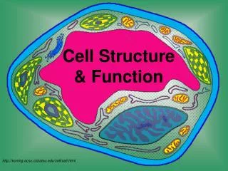
CELLS: THE LIVING UNITS OVERVIEW OF THE CELLULAR BASIS OF LIFE
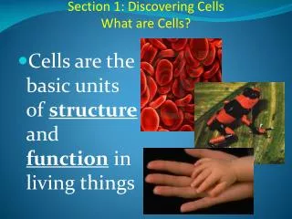
Cell Theory • Cells are the basic structural and functional units of life • The activity of an organism depends on both the individual and the collective activities of its cells • The biochemical activities of a cell are dictated by their organelles • The continuity of life has a cellular basis
Characteristics of Cells • All cells are composed primarily of carbon, hydrogen, nitrogen, and oxygen
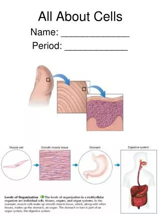
Characteristics of Cells • All cells have the same basic parts and some common functions • A generalized human cell contains the plasma membrane, the cytoplasm, and the nucleus
Plasma Membrane: Structure • Plasma membrane (cell membrane) defines the extent of the cell, separating two of the body’s major fluid compartments: • Intracellular fluid within cells • Extracellular fluid outside cells
The Fluid Mosaic Model • Plasma membrane is composed of a double layer of phospholipids embedded with small amounts of cholesterol and proteins dispersed in it • The phospolipid bilayer is composed of two layers of phospholipids lying tail to tail: • Polar headis charged and hydrophilic (hydro=water, philic=loving) • Exposed to water inside (intracellular) and outside (extracellular) the cell • Attracted to water • Nonpolar tailis made of two fatty acid chains and is hydrophobic (phobia=hating) • Avoid water • Line up in the center of the membrane
The Fluid Mosaic Model • All biological membranes share a common structure: • They are composed of two parallel sheets of phospholipid molecules lying tail to tail, with their polar heads exposed to water inside and outside • This self-orienting property of phospholipids encourages biological membranes to self-assemble into closed, generally spherical, structures and to reseal themselves quickly when torn
The Fluid Mosaic Model • The inward-facing and outward-facing surfaces of the plasma membrane differ in the kinds and amounts of lipids they contain: • The majority of membrane phospholipids are unsaturated (like phosphatidyl choline), a condition which kinks their tails (increasing the space between them) and increases fluidity • Glycolipids, phospholipids with attached sugar groups, are found only in the outer membrane (5% of membrane) • Sugar group makes that end of the glycolipid molecule polar, whereas the fatty acid tails are nonpolar • Cholesterol (20% of membrane) stabilizes the lipid membrane by wedging its platelike hydrocarbon rings between the phospholipid tails and restraining movement of the phospholipids • Lipid rafts (20%), dynamic assemblies of saturated phospholipids (which pack together tightly) associated with unique lipids called sphinolipids and lots of cholesterol are also found only in the outer membrane • More stable and orderly and less fluid than the rest of the membrane • Include or exclude specific proteins to various extents • Assumed to function in cell signaling
The Fluid Mosaic Model • Two distinct populations of membrane proteins: • Integral • Peripheral
Functions of Membrane Proteins • Proteins make up about 50% of the plasma membrane by mass and are responsible for most of the specialized membrane functions • Transport • Enzymatic activity • Receptors for signal transduction • Intercellular joining • Cell-cell recognition • Attachment to the cytoskeleton and extracellular matrix (ECM)
Functions of Membrane Proteins • Transport: • (a):A protein that spans the membrane may provide a hydrophilic channel across the membrane that is selective for a particular solute • (b):Some transport proteins hydrolyze ATP as an energy source to actively pump substances across the membrane
Functions of Membrane Proteins • Enzymatic activity: • A protein built into the membrane may be an enzyme with its active site exposed to substances in the adjacent solution • In some cases, several enzymes in a membrane act as a team that catalyzes sequential steps of a metabolic pathway as indicated (right to left) here
Functions of Membrane Proteins • Receptors for signal transduction: • A membrane protein exposed to the outside of the cell may have a binding site with a specific shape that fits the shape of a chemical messenger, such as a hormone • The external signal may cause a conformational change in the protein that initiates a chain of chemical reactions in the cell
Functions of Membrane Proteins • Intercellular joining: • Membrane proteins of adjacent cellsmay be hooked together in various kinds of intercellular junctions • Some membrane proteins (CAMs) of this group provide temporary binding sites that guide cell migration and offer cell-to-cell interactions
Functions of Membrane Proteins • Cell-Cell recognition: • Some glycoproteins (proteins bonded to short chains of sugars) serve as identification tags that are specifically recognized by other cells
Functions of Membrane Proteins • Attachment to the cytoskeleton and extracellular matrix (ECM): • Elements of the cytoskeleton (cell’s internal supports) and the extracellular matrix (ECM) may be anchored to membrane proteins, which help maintain cell shape and fix the location of certain membrane proteins • Others play a role in cell movement or bond adjacent cells together
The Fluid Mosaic Model • There are two distinct types of membranes proteins: • Integral • Peripheral
Integral Membrane Proteins • Firmly inserted into the plasma membrane • Some protrude from one membrane face only, BUT most are transmembrane proteins that span the entire width of the membrane and protrude on BOTH sides • All have BOTH hydrophobic and hydrophilic regions: • This allows them to interact BOTH with the nonpolar lipid tails buried in the membrane and with water inside and outside the cell
Integral Membrane Proteins • Mainly involved in transport: • Some cluster together to form channels, or pores, through which small, water-soluble molecules or ions can move, thus bypassing the lipid part of the membrane • Some act as carriers that bind to a substance and then move it through the membrane • Some are receptors for hormones or other chemical messengers and relay messages to the cell interior (process called signal transduction)
Peripheral Membrane Proteins • Are not embedded in the lipid of the plasma membrane, but attach rather loosely to integral proteins or membrane phospholipids and are easily removed without disrupting the membranes • Include a network of filaments that helps support the membrane from its cytoplasmic side • Peripheral proteins may function as enzymes or in mechanical functions of the cell, such as changing cell shape during cell division and muscle cell contraction, or linking cells together
Peripheral Membrane Proteins • Many of the proteins that abut the extracellular space are: • Glycoproteins that have branching sugar groups • The term glycocalyx (sugar coated) is used to describe the fuzzy, sticky carbohydrate-rich area at the cell surface • The glycocalyx is enriched BOTH by glycolipids and by glycoproteins secreted by the cells that cling to its surface
Glycocalyx • Because every cell type has a different pattern of sugars in its glycocalyx, the glycocalyx provides highly specific biological markers by which approaching cells recognize each other; • Example: • A sperm recognizes an ovum (egg cell) by the ovum’s unique glycocalyx • Cells of the immune system identify a bacterium by binding to certain membrane glycoproteins in the bacterial glycocalyx • Definite changes in the glycocalyx occur in a cell that is becoming cancerous • A cancer cell’s glycocalyx may change almost continuously, allowing it to keep ahead of immune system recognition mechanisms and avoid destruction
Fluid Mosaic Model • The plasma membrane is a dynamic fluid structure that is in constant flux • Its consistency is like that of olive oil • The lipid molecules of the bilayer move freely from side to side, parallel to the membrane surface, but their polar-nonpolar interactions prevent them from flip-flopping or moving from one leaflet (half of the bilayer) to the other • Some of the proteins float freely • Others, particularly the peripheral proteins, are restricted in their environments because they are “tethered” to intercellular structures that make up the cytoskeleton
Specialization of the Plasma MembraneMicrovilli • Microvilli are fingerlike extensions of the plasma membrane that increase the surface area of the cell • Most often found on the surface of absorptive cells such as intestinal and kidney tubule cells • Have a core of actin filaments • A contractile protein, BUT in microvilli it appears to function as a mechanical stiffener
Specialization of the Plasma MembraneMembrane Junctions • Three factors act to bind cells together: • 1. Glycoproteins in the glycocalyx act as an adhesive • 2. Wavy contours of the membranes of adjacent cells fit together in a tongue-and-groove fashion • 3. Special membrane junctions are formed
Specialization of the Plasma MembraneTight Junctions • Type of membrane junction in which integral proteins on adjacent cells fuse together to form an impermeable junction that encircles the cell • Prevent molecules from passing through the extracellular space between adjacent cells • Example: • Tight junctions between epithelial cells lining the digestive tract keep digestive enzymes and microorganisms in the intestine from seeping into the bloodstream
Specialization of the Plasma MembraneDesmosomes • Desmosomes: binding bodies • Are mechanical couplings that are scattered like rivets along the sides of adjoining cells that prevent their separation • Abundant in tissues subjected to great mechanical stress: • Skin • Heart muscles