Download
1 / 54
740 likes | 1.41k Views
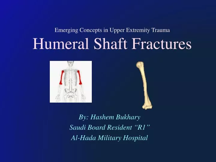
Emerging Concepts in Upper Extremity Trauma Humeral Shaft Fractures. By: Hashem Bukhary Saudi Board Resident “ R1 ” Al-Hada Military Hospital. Humeral Shaft Fractures 3-5% of all fractures Bimodal age distribution: [ High -energy inj. in Healthy Young M VS

E N D
Emerging Concepts in Upper Extremity TraumaHumeral Shaft Fractures By: Hashem Bukhary Saudi Board Resident “R1” Al-Hada Military Hospital
Humeral Shaft Fractures 3-5% of all fractures Bimodal age distribution: [ High-energy inj. in HealthyYoungM VS Low-energy inj. in ElderlyOsteopenic F ] In Children Fracturesof the humerus are Uncommon So in < 3 years of age the possibility of child abuse should be considered .
Humeral Shaft Fractures Generally Nonsurgical management has been supported as the preferred treatment: high union rates + minimal functional deficit “rich vascular supply of muscles”
Anatomy Humeral Shaft : Upp. border of insertion of the Pectoralis Major “Prox” Upp. border of Supracondylar ridge “Dist”
Anatomy Muscles • Insertion for Pectoralis major Deltoid Coracobrachialis • Origin for Brachialis Triceps Brachioradialis
Anatomy • Fracture alignment : Determined by location of the fracture relative to the major muscle attachments “Deforming Forces”
Anatomy • Deforming Forces: • Pectoralis Major • Deltoid attch
Anatomy Radial Nr. “Post. Cord” : [ Triceps Mus. + All 12 Mus. that Extend Forearm ] Courses along Spiral groove
Humeral Shaft Fractures Mechanism of injury Direct or Indirect blow to the arm. Violent muscle contraction to a shaft in an elderly patient with a Pathological Condition “e.g. Metas.”
Humeral Shaft Fractures Classification based on fracture Descriptors. AO Classification
Humeral Shaft Fractures Classification based on fracture Descriptors : Location Pattern ”simple, wedge, complex “ Low-energy vs. high-energy Open / Closed Injury Classifications
Clinical features C/C of long bone # : Pain , Bruise , Swelling, Deformity. Neurovascular must be evaluated & docum (Pre&Post) ”esp: Radial nerve function before and after Rx. Best to be by assessing: Active exte. Metacarpophalangeal Joints WHY? Wrist Ext. can be misleading by (extensor carpi radialis longus) may supplied by a branch arising proximal to the injury.
Humeral Shaft Fractures Imaging: AP & Lat with include Joint above and below.
Humeral Shaft Fractures Management : Conservative: Splint & Brace Surgical: ORIF & CRIF
Humeral Shaft Fractures Conservative: Require neither perfect reduction nor immobiliz. Arm Weight + External cast alignment “Hanging cast”: With the elbow flexed 90 degrees apply a cast from shoulder to elbow, and the forearm section is suspended by a sling around the patient’s neck.
HumeralShaftFractures Conservative:
Humeral Shaft Fractures Conservative: Replaced after 2–3 weeks by a short functional polypropylene brace which is worn for a further 6 weeks. P.T. should begin ASAP: Pendulum exercises of the shoulder are begun within a week & active elbow flexion as soon as comfortable. Active abduction is postponed until the fracture has united (= 6 weeks for spiral fractures & twice as long for other). Only a sling is needed until the fracture is consolidated.
Humeral Shaft Fractures Conservative:Arm Brace
Humeral Shaft Fractures Conservative: What is Acceptable Alignment? < 20° anterior/ post angulation < 30° varus/valgus angulation < 3 cm shortening
Humeral Shaft Fractures Surgical: We should need to remember: The majority of humeral # unite with non-operative. Complic. rate after Intern. Fix. of the humerus is HIGH. There is NO good evidence that the Union rate is higher with fixation , ( But the rate may be lower if there is distraction with nailing or periosteal plating ).
Humeral Shaft Fractures Surgical:Well defined Indications for surgery: An Open fracture Sever Soft .Tiss inj Pathological fracture Vascular injury requiring repair Brachial Plexus Inj. Ipsilateral operative forearm fracture Floating Elbow (Simultaneous Unstable. Hum. + Forearm #) Associated intra-articular extension of the #
Humeral Shaft Fractures Surgical: Fixation can be achieved with : Compression plate and screws. Interlocking intramedullary nail. External fixator.
Humeral Shaft Fractures Surgical: Compression plate and screws: Considered the preferred mode of Fix : Reliable union e.g ( Elderly shaft fractures) Lower reoperation Less effect on adjacent joints Construct to the personality of the fracture: Simple fractures receive compression plating Comminuted segments can undergo bridge plating techniques
Humeral Shaft Fractures Surgical: Compression plate and screws Transverse mid-diaphyseal # High risk of nonunion esp. with conservative management due to distraction forces + gapping across the fracture
Humeral Shaft Fractures Surgical: Compression plate and screws Comminuted mid-diaphyseal #“ +/- radial nerve palsy” Posterior bridge plate using a postero-lateral peri-articular distal humerus plate.
Humeral Shaft Fractures Surgical: Compression plate and screws Long plates create relativestability constructs in comminuted # + micro motion to promote healing. Fractures with poor bone quality can be augmented with locking or hybrid constructs. Humeral shaft # w/ bone loss can be shortened up to 2 cm without affecting muscle strength and improves healing by allowing cortical apposition and compression.
Humeral Shaft Fractures Surgical: Minimally invasive plating osteosynthesis (MIPO) # can reduced w/ closed manipulation indicated in shaft fractures at least 6 cm below the surgical neck and 6 cm above the coronoid fossa to accommodate 6 cortices. Cosmeticincisions. Less soft tissue dissection. Preserved biologic environment at the # Protection of the radial nerve.
Humeral Shaft Fractures Surgical: (MIPO) Forearm is supinated app. Prox. and distal incisions for the MIPO technique for anterior - plating. The proximal incision uses the interval between the bicep and deltoid. The distal incision goes lateral to the bicep and splits the brachialis.
Humeral Shaft Fractures Surgical: Interlocking intramedullary nail: IMN (Intramedullary Nails) offers other mechanical advantages over plates and screws IMN can be inserted without direct fracture exposure minimiz soft-tissue scarring due Smaller incis. &Maintenance of fracture hematoma
Humeral Shaft Fractures Surgical: Interlocking intramedullary nail: IN IMN the implant is Closer to the mechanical axisthan a plate, they are subject to smaller bending loads than plates and are less likely to fail . Indications include : # w/ ass. soft tissue injury preventing an open approach. Pathologic & Impedingfractures Elderly shaft fractures& Extreme osteopenia
Humeral Shaft Fractures Surgical: Intramedullary nail: Use to be Less in the upper extremity # ; Why ? More Stiffness from capsular adhesion, Nonunion, More pain.
Humeral Shaft Fractures Surgical: Intramedullary nail: The New Modern Nails today are larger, more rigid, anatomically- contoured, and statically locked with multiple proximal and distal interlocks : Share load with the bone + Avoid stress shielding. Need Minimal reaming which shown to reduce nonunion and implant failure.
Humeral Shaft Fractures Plating vs Nailing: New Studies showed both are equivalent in union & functional outcomes (modern nails vs compression plate) A Meta-analysis establishing higher shoulder impingement and reoperation with humeral nailing was recently updated and showed NO statistical difference in total complications, nonunion, infection, nerve palsy, or reoperation between plates and nails. A retrospective review of 91 subjects demonstrated NO difference in fracture union and reoperation.
Humeral Shaft Fractures Surgical: External fixator: Severe open fractures with extensive soft-tissue injury or bone loss Associated burns Infected nonunions Humeral shaft fracture with neurovascular injury
Humeral Shaft Fractures Surgical: External fixator: When pt. is hemodynamically stable conversion to an internal fixation + associated debridement + wound coverage in injuries with soft tissue compromise. THEN : Conversion to Plate was thought to be safe within 2 weeks NO data to suggest a safe time for conversion to an IMN”
Complications Radial Nerve Injury. Nonunion. Joint Stiffness .
Radial Nerve Injury 8-15% of Closed fractures Increased incidence distal 1/3 # Holstein-Lewis fracture : Spiral # of the Distal 1/3 of the hum. shaft commonly ass. w/ neuropraxia of the radial Nr. NeuropraxiaVSNeurotmesis ( Closed # ) ( Open # ) Temp.Minor Compresion or contusion “loss continuty of con.tis. & axon of Nr”
Management Radial Nr. Inj: 1/ Observation: 85-90% of improve with Observation in Close # Spontaneous recovery Start at an average of 7 wks . Full recovery = ( average of 6 months ). Obtain “ElectromyographyEMG”at 3-4 months if still. Polyphasic is a good sign. 1st Regain Wrist Extin. Brachioradialis first to recover.
Management Radial Nr. Inj: 2/ Surgecal Exploration: If: Open fracture with radial nerve palsy. Closed fracture that fails to improve over ~ 3-6 months Sharp P wave or Fibrillations (denervation) seen at 3-4 months on EMG Denervation Debrid. Wound + Explor Nr. + Fix # With +/- ( Nr. Repair/graft/ Tendon transfer)
Nonunion (0% to 15%) Mostly Proximal and Distal aspects Hum. Patient factors : Poor nutrition, medical comorbidities, alcohol use, and poor compliance, Old , Steroid & anti coagulant. Fracture factors : Open fractures, midshaft transverse or short oblique & comminution #, unstable fixation, significant bone gaps.
Nonunion The general guideline is 3 months for a delayed union and 6 months to declare a non-union. Infection should always be assessed by : ( Preop. Lab + Multiple C/S # at time of oper. Deb ) If infection confirmed staged Rx: Removal implant Debridement + bone recanalization + antibiotic cement spacer + temporary stabilization IV . Abx specific to the cultured organism ORIF with abundant Autograft is preferred .
Nonunion Nonunions associated with significant Bone Loss due to High-energy open injuries ass. w/ soft tissue and bone defects : May require more elaborate reconstructive efforts Vascularized fibular transfers, I.M. fibular grafting, Even Ilizarov techniques may be needed.
Nonunion Non-Union w/ segmental bone loss from an initial high-energy wound with multi. Failure: Reconst. debridement, culture, Abx, spacer with Temp. Fix After 6 weeks of IV Abx Reconst. w/ titanium mesh spine cage filled with bone graft to rebuild bone stock + supp. IMN