Download
1 / 34
430 likes | 572 Views
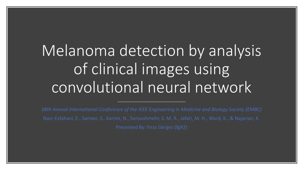
Melanoma detection by analysis of clinical images using convolutional neural network. 38th Annual International Conference of the IEEE Engineering in Medicine and Biology Society (EMBC) Nasr-Esfahani, E., Samavi, S., Karimi, N., Soroushmehr, S. M. R., Jafari, M. H., Ward, K., & Najarian, K.

E N D
Melanoma detection by analysis of clinical images using convolutional neural network 38th Annual International Conference of the IEEE Engineering in Medicine and Biology Society (EMBC) Nasr-Esfahani, E., Samavi, S., Karimi, N., Soroushmehr, S. M. R., Jafari, M. H., Ward, K., & Najarian, K. Presented By: Firas Gerges (fg92)
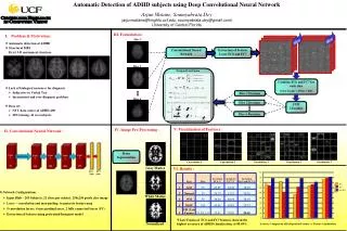
Aim The aim of this study is to detect the existence of melanoma lesions using deep learning
Computer-Aided Methods • Previous studies used image analysis techniques to produce the ABCD features. • Use ML technique to classify a cancerous mole based on the extracted features
Pre-Processing • Three main stages of image handling exists: • Illumination Correction • Segmentation • Filtering
Pre-Processing: Illumination Correction • Illumination effects are detected as sharp changes in HSV color space in terms of saturation and value channels • Some range of gradients (changes in colors and saturation) are excluded from the image to disregard the lightning effects.
Pre-Processing: Filtering • In this step, a Gaussian filter is applied on the normal regions of the skin • This is done to smooth the area outside the lesion, hence reducing this area effect on the classification
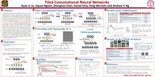
CNN Architecture • The network consists of a set of layers • A layer can be: • Convolve Layer • Pooling Layer • Last layer: fully connected layer
CNN Architecture • Conv. Layers uses a set of kernels to filter the input image • Each Conv. layer is followed by a pooling layer • The pooling layer reduces the size of the constructed feature map • This happens to recognize general patterns in the images. • These general patterns are observable in resized image.
Comparing results • Results were benchmarked against three previous studies done on the same data: • (Zagrouba & Barhoumi, 2004) [24] is one of the first reported work, and used as baseline • (Munteanu & Coocleam, 2009) [13] is an example of used commercial tools • (Giotis et al, 2015) [12] classify cases in a semi-supervised framework
Reference • Nasr-Esfahani, E., Samavi, S., Karimi, N., Soroushmehr, S. M. R., Jafari, M. H., Ward, K., & Najarian, K. (2016, August). Melanoma detection by analysis of clinical images using convolutional neural network. In 2016 38th Annual International Conference of the IEEE Engineering in Medicine and Biology Society (EMBC) (pp. 1373-1376). IEEE. • E. Zagrouba and W. Barhoumi, “A preliminary approach for the automated recognition of malignant melanoma, ” Image Analysis & Stereology, vol. 23, no. 2, pp. 121-135, 2004. • C. Munteanu and S. Cooclea, “Spotmole – melanoma control system,” 2009. Available: http://www.spotmole.com/ • I. Giotis, N. Molders, S. Land, M. Biehl, M. F. Jonkman and N. Petkov, “MED-NODE: A computer-assisted melanoma diagnosis system using non-dermoscopic images,” Expert Systems with Applications, Elsevier, vol. 42, no. 19, pp. 6578-6585, 2015.