Download
1 / 3
30 likes | 34 Views
Mucormycosis is a rare invasive opportunistic fungal infection with high morbidity and mortality, with Intraparenchymal Hemorrhage (ICH) being one of the less common complications cited in available literature. In this case report, we present a case of a 65 year old Filipino diabetic male with non-small cell lung cancer and recent history of steroid use who presented with headache. This patient developed proptosis, chemosis, and purulent discharge from the right eye, and eventually had sudden onset decrease in sensorium on the 6th day of admission. CT scan showed findings of ICH on the right f

E N D
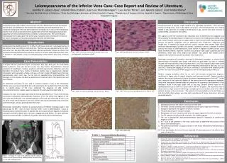
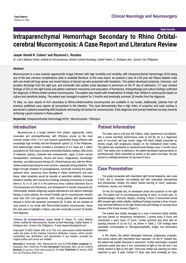
Clinical Neurology and Neurosurgery Case Report Volume 5:1, 2022 Open Access Intraparenchymal Hemorrhage Secondary to Rhino Orbital- cerebral Mucormycosis: A Case Report and Literature Review Jasper Gerald R. Cubias* and Raymond L. Rosales St. Luke’s Medical Center, Institute for Neurosciences, Section of Adult Neurology, Capital Towers, E. Rodriguez Ave., Quezon City, Philippines Abstract Mucormycosis is a rare invasive opportunistic fungal infection with high morbidity and mortality, with Intraparenchymal Hemorrhage (ICH) being one of the less common complications cited in available literature. In this case report, we present a case of a 65 year old Filipino diabetic male with non-small cell lung cancer and recent history of steroid use who presented with headache. This patient developed proptosis, chemosis, and purulent discharge from the right eye, and eventually had sudden onset decrease in sensorium on the 6th day of admission. CT scan showed findings of ICH on the right frontal and patient underwent craniotomy and evacuation of hematoma. Histopathology and culture findings confirmed the diagnosis of Rhino-orbital-cerebral mucormycosis. The patient was treated with Amphotericin B initially then shifted to voriconazole based on culture and sensitivity testing. The patient was managed in-patient for 3 months and eventually survived 18 months from the diagnosis. To date, no case reports of ICH secondary to Rhino-orbital-cerebral mucormycosis are available in our locale. Additionally, patients from all previous published case reports all succumbed to the infection. This case demonstrates that a high index of suspicion and early workup is warranted in patients presenting with headache and having risk factors for mucormycosis. Early diagnosis and prompt treatment are keys towards achieving a good outcome in these patients. Keywords: Intraparenchymal Hemorrhage (ICH) • Mucormycosis • Rhizopus Introduction Mucormycosis is a fungal infection from phylum zygomycota, orders mucorales and entomophthorales, with Rhizopus oryzae as the most common isolate (70%). It is a rare invasive opportunistic fungal infection with exceedingly high mortality and few therapeutic options [1]. In the Philippines, local epidemiologic studies revealed a prevalence of 0.2 cases per 1 million populations [2]. Risk factors include uncontrolled diabetes mellitus, other forms of metabolic acidosis, treatment with corticosteroids, organ or bone marrow transplantation, neutropenia, trauma and burns, malignancies, hematologic disorders, and deferoxamine therapy [3]. Clinical features vary, with the Rhino- orbital-cerebral type being the most common, especially among diabetics. This begins through inhalation of sporangiospores, eventually involving the palate, sphenoid sinus, cavernous sinus (leading to orbital involvement) and brain tissue. Initial symptoms would be sinusitis or periorbital cellulitis. Extensive infections manifest with cranial nerve findings including involvement of Cranial Nerve II, III, IV, VI, and VI in the cavernous sinus; cerebral infarctions due to ICA encasement and thrombosis, and development of mycotic aneurysms [4]. Unfortunately, despite disfiguring surgical debridement and adjunct antifungal therapy in some patients, the overall mortality rate for mucormycosis remains more than 50% and approaches 100% among patients with disseminated disease or those with persistent neutropenia [5]. To date, we are unaware of case reports in our locale with Rhino-orbital-cerebral mucormycosis, hence this work aims to highlight a Filipino case who eventually survived 18 months from diagnosis. Patient Information The index case is a 65 year old, Filipino, male, hypertensive and diabetic, with a cancer karnofsky performance scale of 100 [6]. He is a diagnosed case of non-small cell lung cancer, stage IVB which initially presented as chronic cough, with progressive disease on the mediastinal lymph nodes. The patient was maintained on pembrolizumab therapy every 3 months since 2017.Two weeks prior to admission, the patient developed hypersensitivity to pembrolizumab, which presented as rashes on the palms and soles. He was started on methylprednisolone 16 mg every 8 hours. Case Presentation The patient presented with intermittent right frontal headache, pain scale 1-3/10, dull in character non-radiating and with associated phonophobia and photophobia. Initially, the patient denied blurring of vision, weakness, numbness, nausea, and vomiting. On the 3rd hospital day, he developed ptosis and proptosis on the right eye. The patient had no accompanying blurring of vision, no ocular pain, no ophthalmoplegia, chemosis, diplopia, vomiting, or fever. A noncontrast cranial MRI showed right orbital cellulitis. Additional findings included a sliver of extra- axial restricted diffusion on the right frontal area and findings of mucosal sinus disease of the frontal sinuses (Figure 1). The patient was initially managed as a case of bacterial orbital cellulitis and was started on intravenous meropenem, 2 grams every 8 hours and vancomycin 1 gram every 12 hours. Lumbar puncture was done with an opening pressure of 18 cm H2O. As shown in Table 1, CSF findings were essentially unremarkable on meningoencephalitis, fungal, and tuberculous infections. *Address for Correspondence: Jasper Gerald R. Cubias, St. Luke’s Medical Center, Institute for Neurosciences, Section of Adult Neurology, Capital Towers, E. Rodriguez Ave., Quezon City, Philippines; E-mail: jaspercubias@gmail.com Copyright: © 2022 Cubias JGR, et al. This is an open-access article distributed under the terms of the Creative Commons Attribution License, which permits unrestricted use, distribution, and reproduction in any medium, provided the original author and source are credited. Received:12 November, 2021, Manuscript No. jcnn-21-47285; Editor assigned:15 November, 2021, PreQC No. P-47285; Reviewed: 14 November, 2021, QC No. Q-53575; Revised: 12 January, 2022, Manuscript No. R-47285; Published: 18 January, 2022, DOI: 10.37421/jcnn.2022.5.131 In the interim, the patient developed chemosis, progressive proptosis, and scleral edema with intermittent fever episodes. On the 6th hospital day, the patient had sudden decrease in sensorium. Further examination revealed anisocoric pupils who were 4 mm, nonreactive to light on the left and 3 mm briskly reactive to light on the right. There was neither eye opening nor motor response to pain. A plain Cranial CT Scan was done revealing an Intra-
Cubias JGR, et al. Clin Neurol Neurosurg, Volume 5:1, 2022 parenchymal Hemorrhage (ICH) proximately 60 milliliters on the right frontal lobe with intra-ventricular extension and a leftward midline shift of 1.4 cm (Figure 2). The patient underwent an emergency right frontal craniectomy, evacuation of intra-cerebral hematoma, and insertion of External Ventricular Drain (EVD). KOH smear of the organized hemorrhage collected revealed many hyphae with irregular widths and right-angle branching. External ventricular drain aspirates were sent for culture studies which revealed filamentous fungi on histopathology (Figure 3). On further speciation and sensitivity testing, the Figure 3. High power (400 X) H and E stain of EVD aspirate showing filamentous fungi. Table 2. Literature review of case reports of mucormycosis related intraparenchymal hemorrhages. Figure 1. Contrast enhanced MR imaging of the right frontal region. Authors Munoz J, et al. [5] Age/Sex ICH Findings Bilateral Frontoparietal ICH Place of Report Risk Factors High dose steroid use Myelodysplastic syndrome acute renal failure Leukemia allogeneic bone marrow transplantation Outcome Table 1. Lumbar puncture results. Died 48/M Detroit, USA Test Opening Pressure Colour Result Test Result Left Parietooccipital ICH Scully MA, et al. [8] New York, USA Died 77/M 10 cm H20 18 cm H2O Closing pressure Clear CALAS Negative Died on postop Day 36 Takahashi S, et al. [10] Meningoencephalitis Panel *Cytomegalovirus; Cryptococcus neoformans/gatti; E. Coli K1; Enterovirus; H. Influenzae; HHV6, HSV-1, HSV-2, Listeriamonocytogenes; N. Meningitidis, Streptococcus agalactiae; VZV, Streptococcus pneumonia; Human parechovirus 54/M Left frontal ICH Tokyo, Japan Total cell count Left Frontal intracerebral hematoma with subarachnoid haemorrhage 12 Negative Uncontrolled diabetes mellitus Koc Z, et al. [11] Adana, Turkey Died 47/M Chronic neutrophilic leukemia prednisone use No Died at hospital Day 19 Right Yasui H, et al. [9] RBC 4 gram stain microorganisms seen No hyphal elements seen No growth after 3 days Negative Sapporo, Japan 67/M frontotemporal ICH 8 (all WBC KOH Died 6 hours after admission Rocha O, et al. [7] *full article not available Hyvernat H, et al. [6] **full article and abstract not available lymphocytes) Gastric Carcinoma Parietooccipital hematoma 38/F Lisbon, Portugal Protein 50 Culture Glucose 61 (Ratio 0.7) MTB-PCR and AFB _ _ _ _ Survived* organism isolate turned out to be Rhizopusmicrosporus complex, consistent with the diagnosis of mucromycosis. The patient was admitted to the neurocritical care unit and was started on Amphotericin B 300 mg IV every 24 hours for 33 days, with voriconazole 240 mg every 24 hours which was continued for 63 days. Follow-up and outcomes The patient was discharged after 3 months of admission his hospital course complicated by pneumonia and kidney injury, but eventually had improvement of neurologic status. Patient also had a tracheostomy and Percutaneous Endoscopic Gastrostomy (PEG) as he was chronic home care. Currently, 18 months henceforth, the patient has been sent back home to his province with his family. On follow up via telemedicine, patient is dependent on his caregivers for feeding, bathing, and other activities of daily living. He has spontaneous eye opening, but with no regard, recognition, or speech. Both eyeballs can cross midline, with a 3 mm briskly reactive pupil in the left. He has intact mouth and tongue movements. All his extremities are plegic and spastic, with a modified Ashworth scale of 3 [7]. He is hyporeflexive on all extremities Figure 2. Plain Cranial CT Scan showing right front parietal intra-parenchymal haemorrhage. Page 2 of 3
Cubias JGR, et al. Clin Neurol Neurosurg, Volume 5:1, 2022 and an absent Babinski sign. He regularly undergoes physical therapy and stretching sessions. He is still maintained on levetiracetam 500 mg 2x/day and citicholine 500 mg 2x/day. At best, he is able to do wheelchair rides when assisted by his caregivers. In conclusion, patients with traditional risk factors for mucormycosis such as malignancy and steroid use such as that seen in our patient should have a high index of suspicion and warrant early workup. Prompt diagnosis and treatment is the key in ensuring a good outcome in these patients. Discussion References Mucormycosis is being increasingly recognized in immune compromised hosts and carries a poor prognosis. Early recognition and treatment are critical in order to improve clinical outcomes [8]. In extensive infections, patients manifest with cranial nerve findings, cerebral infarctions, or even more rarely, intra-parenchymal hemorrhage [9]. Underlying pathophysiology is likely due to mucor fungi’s high affinity for blood vessels, particularly for the elastic membrane, making the patient more prone to vascular invasion. In a previous case report, intraoperative findings and a contrast CT scan revealed an embolized vein opening up the venous occlusion as a potential pathogenesis for intra-parenchymal hemorrhages. 1. Lewis R. E and Kontoyiannis D. P. “Epidemiology and treatment of mucormycosis.” Future Microbiol (2013) 8: 1163-1175. 2. Batac M. C. R and Denning D. “Serious fungal infections in the Philippines.” Eur. J. Clin. Microbiol. Infect. Dis 36 (2017): 937-941. 3. Spellberg B, Edwards J and Ibrahim A. “Novel perspectives on mucormycosis: pathophysiology, presentation, and management.” Clin. Microbiol. Rev (2005) 18: 556-569. 4. Vaughan C, Bartolo A, Vallabh N and Leong SC. “A meta-analysis of survival factors in rhino-orbital-cerebral mucormycosis-has anything changed in the past 20 year?” Clin. Otolaryngol (2018) 43: 1454-1464. Definitive diagnosis of Mucormycosis requires identification of non-septate irregular hyphae that branch at right angles in tissue specimens, which was seen in this patient. The standard treatment includes liposomal Amphotericin B, management of underlying conditions such as immune system deficiency, and aggressive surgical debridement of necrotic tissues [10]. 5. Munoz J, Hughes A and Guo Y. “Mucormycosis-associated intracranial hemorrhage.” Blood Coag & Fibri (2013) 24: 100-101. 6. Hyvernat H, P de Swardt, M Gari-Toussaint and S Castillo-Ros, et al. “Rhino- cerebral mucormycosis complicated by intracerebral hemorrhage with favorable outcome.” PubMed (2000) 29: 1762. Ninety day mortality is in excess of 50%, particularly in the population of patients with rhino cerebral mucormycoses [11]. A literature search in Pubmed using the terms “intraparenchymal hemorrhage” and “mucormycosis” and (case report or case series) revealed 7 case reports of mucormycosis associated intraparenchymal hemorrhages as detailed in Table 2. 7. Oliviera Rocha, Victor Augusto and María Adília Costa Ferreira. “Brain hematoma caused by mucormycosis.” Revista de Neurología (2001) 10: 951. 8. Scully M. A, Yeaney G. A, Compton M. L and Berg, M. J. “SWAN MRI revealing multiple microhemorrhages secondary to septic emboli from mucormycosis.” Neurol (2012) 79: 1932-1933. Conclusion 9. Yasui H, Adachi Y, Ishii Y and Kato Y. “Mucormycosis as an etiology of cerebral hemorrhage in patients with chronic neutrophilic leukemia.” Am. J. Med (2003) 115: 674-676. The above literature review showed that 6 out of 7 available case reports had a fatal outcome. Ages ranged from 38 year-77 years old and traditional risk factors stated above were all identified in the patient population. Due to the low prevalence of this disease, especially in the subset of patients who develop ICH from its ROCM variant, no large-scale or randomized studies have yet been documented. In this case report, we presented a rare phenomenon of a Filipino patient with lung malignancy and steroid use, which developed intra- parenchymal hemorrhage secondary to rhino-orbital-cerebral mucormycosis and achieved survival 18 months after admission. 10. Takahashi S, Horiguchi T, Mikami S and Kitamura Y et al. “Subcortical intracerebral hemorrhage caused by mucormycosis in a patient with a history of bone-marrow transplantation.” J. Str. Cerebro. Dis (2009) 18: 405-406. 11. Koc Z, Koc F, Yerdelen D and Ozdogu H. “Rhino-orbital-cerebral mucormycosis With different cerebral involvements: infarct, hemorrhage, And ophthalmoplegia.” Int. J. Neurosci (2007) 117: 1677-1690. How to cite this article: Cubias, Jasper Gerald R. and Raymond L. Rosales. “Intraparenchymal Hemorrhage Secondary to Rhino Orbital-cerebral Mucormycosis: A Case Report and Literature Review.” Clin Neurol Neurosurg 5(2022): 131 Page 3 of 3