Download
1 / 52
550 likes | 867 Views
Management of Patients With Musculoskeletal Disorders. Acute low Back pain:. Causes: A cut lumbosacral strain, unstable lumbosacral ligaments and weak muscles, osteoarithritis of the spine, spinal stenosis, intervertebral disk problems, and unequal leg length.

E N D
Acute low Back pain: • Causes: • A cut lumbosacral strain, • unstable lumbosacral ligaments and weak muscles, • osteoarithritis of the spine, • spinal stenosis, intervertebral disk problems, • and unequal leg length. • Other causes include kidney diorders, pelvic problems, retroperitoneal tumors, abdominal aneurysims, obesity and stress. • L4-L5 and L5-S1 has the greatest degenrative changes
Clinical manifestations: • A cute or chronic back pain lasting more than 3 months without improvement) • Fatique • Pain radiating down the leg ( radiculopathy; Sciatica) • Patient’s gait, spinal mobility, reflexes, leg length, leg motor strength and sensory perception may altered
Cont… • Assessment and diagnostic findings: • Focused history and physical examination ( reflexes, sensory impairment, straight leg raising, muscle strength • X-ray of the spine, CT scan, MRI • Bone scan and blood study • Myelogram and dicogram • Electromyogram and nerve conduction studies • Medical management: Analgesia, rest, stress reduction and relaxation • Review the Nursing process
Nursing Process: The Care of the Patient with Low Back Pain—Assessment • Detailed description of the pain including severity, duration, characteristics, radiation, associated symptoms such as leg weakness, description of how the pain occurred, and how the pain has been managed by the patient • Work and recreational activities • Effect of pain and/or movement limitation on lifestyle and ADLs • Assess posture, position changes, and gait • Physical exam: spinal curvature, back and limb symmetry, movement ability, DTRs, sensation, and muscle strength • If obese, complete a nutritional assessment
Nursing Process: The Care of the Patient with Low Back Pain—Diagnoses • Acute pain • Impaired physical mobility • Risk for situational low self-esteem • Imbalanced nutrition
Nursing Process: The Care of the Patient with Low Back Pain—Planning • Major goals may include relief of pain, improved physical mobility, use of back conservation techniques and proper body mechanics, improved self-esteem, and weight reduction.
Interventions • Pain management • Exercise • Body mechanics • Work modifications • Stress reduction • Health promotion; activities to promote a healthy back • Dietary plan and encouragement of weight reduction
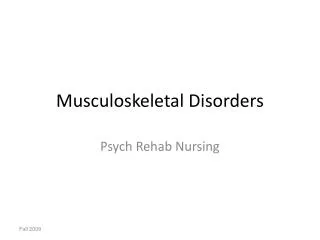
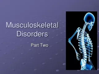
Osteoporosis • Affects approximately 40 million people over the age of 50 in the United States. • Normal homeostatic bone turnover is altered and the rate of bone resorption is greater than the rate of bone formation, resulting in loss of total bone mass. • Bone becomes porous, brittle, and fragile, and break easily under stress • Frequently result in compression fractures of the spine, fractures of the neck or intertrochanteric region of the femur, and Colles’ fractures of the wrist • Risk factors.
Progressive Osteoporosis Bone Loss and Compression Fractures
Risk factors: see chart 68-7 • Assessment and diagnostic findings: • Routine X-ray, and bone sonometer • Lab. Studies: Serum Ca, Serum Ph, urine calcium excretion, ESR • Medical Management: • Adequate balanced diet rich in calcium and Vit D • Regular weight bearing exercise promotes bone formation • Pharmacological therapy: Hormonal replacement therapy ( look for side effect of estrogen and progesterone replacement therapy which result in cancers… thus frequent breast examination is recommended ) • Alendronate alternative to Hormonal replacement therapy: inhibiting osteoclast function and dedcreases bon loss • Calcitonin: suppress bone loss
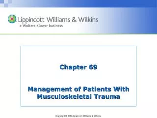
Osteoporosis • Manifestations : • Loss of height • Progressive curvature of spine • Low back pain • Fractures of forearm, spine, hip • Development of “dowager’s hump” • Dorsal kyphosis • Cervical lordosis
Typical Loss of Height Associated with Osteoporosis and Aging
Prevention • Balanced diet high calcium and vitamin D throughout life • Use of calcium supplements to ensure adequate calcium intake—take in divided doses with vitamin C • Regular weight-bearing exercises—walking • Weight training stimulates bone mineral density (BMD)
Pharmacologic Therapy • Biphosphonates • Alendronate: Fosamax • Risedronate: Actonel • Ibandronate: Boniva • Selective estrogen modulators (SERMs): Evista • Cacitonin • Teriparatide: Forteo • Also need adequate amounts of calcium and vitamin D
Injuries of the Musculoskeletal System • Contusion: soft tissue injury produced by blunt force • Pain, swelling, and discoloration: ecchymosis • Strain: Pulled muscle-injury to the musculcoteninous unit • Pain, edema, muscle spasm, ecchymosis, and loss of function are on a continuum graded 1st , 2nd, and 3rd degree • Sprain: injury to ligaments and supporting muscle fiber around a joint • Joint is tender and movement is painful, edema, disability and pain increases during the first 2–3 hours • Dislocation: articular surfaces of the joint are not in contact • A traumatic dislocation is an emergency with pain change in contour, axis, and length of the limb and loss of mobility
RICE • Rest • Ice • Compression • Elevation
Common Sports-Related Injuries • Contusions, strains, sprains and dislocations • Tendonitis: inflammation of a tendon by overuse • Meniscal injuries of the knee occur with excessive rotational stress • Traumatic fractures • Stress fractures
Prevention of Sports-Related Injuries • Use of proper equipment; running shoes for runners, wrist guards for skaters, etc. • Effective training and conditioning specific for the person and the sport • Stretching prior to engaging in a sport or exercise has been recommended but may not prevent injury • Changes in activity and stresses should occur gradually • Time to “cool down” • Tune in to the body; be aware of limits and capabilities • Modify activities to minimize injury and promote healing
Occupational-Related Injuries • Common injuries include strains, sprains, contusions, fractures, back injuries, tendonitis, and amputations. • Prevention measures may include personnel training, proper use of equipment, availability of safety and other types of equipment (patient lifting equipment, back belts), correct use of body mechanics, and institutional policies.
Joint Dislocation • articular surfaces of the joint are not in contact • A traumatic dislocation is an emergency with pain change in contour, axis, and length of the limb and loss of mobility • Most common • Shoulder • Acromioclavicular joints • Subluxation • Partial dislocation
Types of Fractures • Complete • Incomplete • Closed or simple • Open or compound/complex • Grade I • Grade II • Grade III
Cont… • Grade I: clean wound less than 1 cm • Grade II: larger wound without extensive soft tissue damage • Grade III: is highly contaminated, has extensive soft tissue damage, and is the most severe
Manifestations of Fracture • Pain • Loss of function • Deformity • Shortening of the extremity • Crepitus • Local swelling and discoloration • Diagnosis by symptoms and x-ray • Patient usually reports an injury to the area
Emergency Management • Immobilize the body part • Splinting: joints distal and proximal to the suspected fracture site must be supported and immobilized • Assess neurovascular status before and after splinting • Open fracture: cover with sterile dressing to prevent contamination • Do not attempt to reduce the fracture
Medical Management • Reduction • Closed • Open • Immobilization: internal or external fixation • Open fractures require treatment to prevent infection • Tetanus prophylaxis, antibiotics, and cleaning and debridement of wound • Closure of the primary wound may be delayed to permit edema, wound drainage, further assessment, and debridement if needed
Three Phases of Fracture Healing • Inflammatory phase • Reparative phase: collagen formation and calcium deposition • Remodeling phase: 1. Excess callus is removed 2. New bone is laid down 3. Fracture site calcifies 4. Bone is reunited
Three Phases of Fracture Healing • Inflammatory phase • Reparative phase: collagen formation and calcium deposition • Remodeling phase: 1. Excess callus is removed 2. New bone is laid down 3. Fracture site calcifies 4. Bone is reunited
Three Phases of Fracture Healing • Inflammatory phase • Reparative phase: collagen formation and calcium deposition • Remodeling phase: 1. Excess callus is removed 2. New bone is laid down 3. Fracture site calcifies 4. Bone is reunited
Complications of Fractures • Factors that affect fracture healing • Shock • Fat embolism • Compartment syndrome • Delayed union and nonunion • Avascular necrosis • Reaction to internal fixation devices • Complex regional pain syndrome (CRPS) • Heterotrophic ossification
Fracture Complications • Early complications: • Shock: Hypovolemic or traumatic shock result from bleeding or from loss of extracellular fluid • Treatment: 1. Restoring blood volume and circulation 2. Releiving patient pain 3. Provide adequate splinting.
Compartment Syndrome • develop when tissue perfusion in the muscles less than that required for tissue viability. Physiology • Entrapment of the blood vessels limits tissue perfusion • Results in edema within the compartment • Edema causes further pressure Early Manifestations • Deep Pain which is not controlled by opioids • Normal or decreased peripheral pulse Later Manifestations • Cyanosis • Paresthesias, paresis • Severe pain
Treatment • Interventions to alleviate pressure • Removal of the cast • Fasciotomy
Bone Healing Stimulator • Refer to fig. 69-6
3. Fat Embolism Syndrome • Pathophysiology • Bone fracture results in a rise of pressure in the bone marrow • Fat globules enter the bloodstream • Combine with platelets • Travel throughout the body • Occluding small blood vessels • Causes tissue ischemia
Fat Embolism Syndrome • Manifestations • Confusion • Changes in level of consciousness • Pulmonary edema • Impaired surfactant production • Atelectasis • ARDS
Fat Embolism Syndrome • Prevention • Early stabilization of long bone fracture. Treatment: • Intubation and mechanical ventilation • Fluid balance • Corticosteroids • Vasoactive medication to support the cardiovascular system and prevent hypotension.
4. Deep Vein Thrombosis • Manifestations • Swelling • Leg pain • Tenderness • Cramping • Some are asymptomatic • Diagnosis • Doppler ultrasound