Download

1 / 42
600 likes | 1.68k Views
Hypersensitivity reactions. The immune system is concerned with protection of the host against foreign antigens, particularly infectious agents. Inappropriate immune response may be: Allergy : exaggerated immune response against environmental antigens.

E N D
The immune system is concerned with protection of the host against foreign antigens, particularly infectious agents. Inappropriate immune response may be: Allergy: exaggerated immune response against environmental antigens. Autoimmunity: misdirected immune response against the host’s own cells.
3. Alloimmunity: immune response directed against beneficial foreign tissues e.g. blood transfusion or organ transplantation.4. Immune deficiency: inability of the immune system to protect the host.
Hypersensitivity Is an altered immunologic response to an antigen that results in tissue damage.
Hypersensitivity reactions • Type I: immediate (Ig E mediated) hypersensitivity • Type II: Tissue specific (cytotoxic) hypersensitivity • Type III: immune-complex mediated hypersensitivity • Type IV: cell mediated or delayed hypersensitivity
Type I hypersensitivity 1). Characteristics 2). Components and cells 3). Mechanism 4). Clinical examples of type I Hypersensitivity 5). Therapy for type I Hypersensitivity
Type I hypersensitivity reactions are the most common forms of allergic reactions especially against environmental agents.
1)Characteristics Occur and resolve quickly Mediated by serum IgE Systemic and regional tissue dysfuntion Genetic predisposition (atopy)
2)Components and cells in Type I hypersensitivity • (Antigen) Allergen: pollen、dust mite、insects etc • selectively activate CD4+Th2 cells and B cells • Antibody:(IgE): • IgE: mainly produced by mucosal B cells in the lamina prapria. • IL-4 is essential to switch B cells to IgE production • Mast cell and basophil • Eosinophil
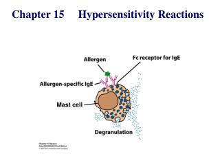
3). Mechanism: * First exposure to allergen Allergen stimulates B lymphocytes to form antibody (IgE type). IgE fixes, by its Fc portion to mast cells and basophils. * Second exposure to the same allergen The antigen fixes directly to IgE (which is already fixed to mast cell) leading to activation and degranulation of mast cells and release of mediators
The biological mediators are: 1. Histamine: Vasodilatation and increased vascular permeability. 2. Leukotrienes: Bronchial smooth muscles contraction 3. Prostaglandin D2: Causes bronchospasm and increased mucin secretion. 4. Platelet activating factor (PAF): platelet aggregation, release of histamine, bronchospasm, increased vascular permeability, and vasodilation 5. Eosinophil chemotactic factor(ECF-A: 6. Bradykinin: Vasodilation
Immediate Phase Allergic Reaction: • Occurs within seconds to minutes of IgE receptor activation (mast cell mediator release) and resolving within an hour • Intense pruritus, edema, erythema
Late Phase Allergic Reaction: • A delayed inflammatory response (peaking at 4-8 hrs and persisting up to 24 hrs) following an intense acute phase reaction • Skin: erythema, induration, burning • Lungs: airway obstruction poorly responsive to bronchodilators • Nose/eyes: erythema, congestion, burning • Histology: infiltration of tissues with eosinophils, neutrophils, basophils, monocytes, and CD4+ T cells as well as tissue destruction, typically in the form of mucosal epithelial cell damage.
Clinical examples of type I hypersensitivity 1. Systemic anaphylaxis: a very dangerous condition Allergic reactions after injection of drugs (penicllin)or serum 2. Respiratory allergic diseases : 1) Allergic asthma:acute response, chronic response 2) Allergic rhinitis, Allergic rhinoconjunctivitis (hay fever 3. Gastrointestinal allergic disease: 4. Skin allergy: Eczema (atopic dermatitis), Acute urticaria
Anaphylaxis * Systemic form of Type I hypersensitivity * Exposure to allergen to which a person is previously sensitized * Allergens: Drugs: penicillin Serum injection : anti-diphtheritic or anti-tetanic serum anesthesia or insect venom * Clinical picture: Shock due to sudden decrease of blood pressure, respiratory distress due to bronchospasm, cyanosis, edema, urticaria * Treatment: corticosteroids injection, epinephrine, antihistamines
Atopy * There is a strong familial predisposition to type I hypersensitvity reaction. * The predisposition is genetically determined * Atopic individuals have higher quantities of IgE antibodies and higher concentration of Fc receptors on mast cells. * The airways and skin are commonly affected. * Allergens : Inhalants: dust mite, pollens, mould spores. Ingestants: milk, egg, fish, chocolate contactants: wool, nylon, animal fur.
Methods of diagnosis 1)History taking for determining the allergen involved 2) Skin tests: Intradermal injection of battery of different allergens A wheal and flare (erythema) develop at the site of allergen to which the person is allergic 3) Determination of total serum IgE level- Radioimmunosorbent test (RIST) 4) Determination of specific IgE levels to the different allergens- Radioallergosorbent test (RAST)
Management 1) Avoidance of specific allergen. 2) Hyposensitization: Minute quantities of the responsible allergen is injected in increasing doses over a long peroid. 3) Drug Therapy: corticosteroids injection, epinephrine, antihistamines
2. Type II Hypersensitivity (Cytotoxic or Cytolytic Reactions) 1. Characteristic features 2. Mechanism of Type II Hypersensitivity 3. Common diseases of Type II Hypersensitivity
1. Characteristic features + Primed IgG or IgM Antigen or hapten on membrane Injury and dysfunction of target cells
Type II Hypersensitivity Reactions:Mechanisms of Tissue Damage An antibody (Ig G or Ig M) reacts with antigen on the cell surface * This antigen may be part of cell membrane or circulating antigen (or hapten) that attaches to cell membrane
* Mechanisms of type II hypersensitivity reactions: • Complement-mediated cell lysis. Complement fixation to antigen antibody complex on cell surface. The activated complement will lead to cell lysis. • Phagocytosis mediated cell lysis. • Phagocytosis is enhanced by the antibody (opsinin) bound to cell antigen leading to opsonization of the target cell
Antibody-dependent cell-mediated cytotoxicity (ADCC): - Antibody coated cells e.g. tumor cells, graft cells or infected cells can be killed by cells possess Fc receptors. • Antibody mediated cellular dysfunction: • The antibody does not destroy the cell but attach certain receptor to either block them (myasthenia gravis) or stimulate them (Grave’s disease).
Clinical examples of type II hypersensitivity reaction: 1) Incompatible blood transfusion:due to ABO incompatibility 2) Rh-incompatability(Haemolytic disease of the newborn) 3) Autoimmune hemolytic anaemia. 4). Autoimmune thrombocytopenic purpura. 5). Myasthenia gravis. 6). Gravis disease. 7). Insulin-resistant diabetes mellitus.
8). Graft rejection cytotoxic reactions: In hyperacute rejection the recipient already has performed antibody against the graft 9). Drug reaction: Penicillin may attach as haptens to RBCs and induce antibodies which are cytotoxic for the cell-drug complex leading to haemolysis Quinine may attach to platelets and the antibodies cause platelets destruction and thrombocytopenic purpura
3. Type III(Immune complex-mediated hypersensitivity reactions. 1. Characteristics 2. Mechanism of type III hypersensitivity 3. Clinical examples of type III hypersensitivity
1. characteristics Free Ag + Primed Ab forming larger immune complexs Deposit in tissue or blood vessel wall complement activation and subsequentInflammation
2. Mechanism of type III hypersensitivity: • Immune complex activate Complement system • Split products-C3a, C4a,C5a. • C3a, C4a, C5a are chemotactic for Neutrophils. • Neutrophils attempt to phagocytose the immune complex which is often unsuccessful because the complexes are bound to a large areas of tissue. • During this attempts, release of large quantities of lysosomal enzymes causing tissue damage and inflammation.
3. Clinical examples of type III hypersensitivity 1. SLE. 2. Polyarteritis nodosa. 3. Post-streptococcal glomerulonephritis. 4. Serum sickness. 5. Arthus reaction.
Arthus reaction • Caused by repeated local exposure to an antigen that reacts with preformed antibody and forms immune complexes in the walls of the local blood vessels. • The symptoms appear within 1 hour after injection and the peak 6-12 hours later. • Lesions include; edema, hemorrhage, clotting and tissue necrosis.
Serum sickness • Is a protoype of systemic immune complex hypersensitivity reaction. • The immune complex circulate in the blood and deposit in blood vessels (vascultitis), joint (arthritis) and kidney (glomerulonephritis) associated with fever, rash and lymphadenopathy. • Serum sickness was initially described as a complication of therapeutic administration of horse serum that contains anti-tetanic Abs. • Serum sickness reactions can be caused by repeated intravenous administration of other antigens such as drugs.
TYPE IV (cell mediated) hypersensitivity • Whereas types I, II and III mediated by antibody, type IV mediated by T lymphocytes (cytotoxic T cells or cytokine-producing Th1 cells). • Develops after 48-72 hrs of second exposure to antigen in a sensitized individual. • Some subpopulations of activated TH cells encounter certain types of antigens, they secrete cytokines that induce a localized inflammatory reaction- delayed-type hypersensitivity (DTH). • The reaction is characterized by large influxes of nonspecific inflammatory cells, macrophages.
A prolonged DTH response leads to destructive inflammatory response with development of granulomatous reaction. • A granuloma develops by continuous activation of macrophages. • Giant cells displace the normal tissue cells, forms palpable nodules, and release high concentrations of lytic enzymes, which destroy surrounding tissue.
PHASES of DTH response • Sensitization phase • Activation of TH cells by Antigen presenting cells (APC) e.g Langerhans cells & Macrophages • Proliferation TH 1 subtype occurs. • Effector phase • TH 1 secrete cytokines , IL2, TNFβ • Recruitment & activation of Macrophages
3. Clinical examples of type IV hypersensitivity 1) Tuberculin test: a skin test for T.B 2) Contact dermatitis : caused by Paint, drug leading to appearance of red rash, papula, water blister, dermatitis 3) Graft rejection after organ transplantation 4) Immune response in local tumor mass
Gell and Coombs classification of hypersensitivity reaction Type Description Time Mechanism, Typical manifestation Type IIgE-mediated 2-3min Ag induce cross-linkage Systemic anaphylaxis hypersensitivity of IgE bound to mast cells Localized anaphylaxis: or basophils with release -Hay fever, Asthma, of vasoactive mediators Hives, Food Allergy Eczema. Type II Ab-mediated 5-8h Ab directed against cell- Blood-transfusion cytotoxic surface Ags mediates cell reactions. hypersensitivity destruction via C activation Erythroblastosis fetalis Autoimmune hemolytic anemia.
Gell and Coombs classification of hypersensitivity reaction Type Description Time Mechanism Typical Manifestations TypeIII Immune complex 2-8h Ag-Ab complexes Localized Arthus -mediated deposited in various reaction hypersensitivity tissues induce C acti- Generalized reactions: vation and an ensuing Serum sickness, inflammatory response Glomerulonephritis Rheumatoid arthritis SLE Delayed reactions Type IV cell-mediated 24-72h Sensitized TDTH cells Contact dermatitis, hypersensitivity release cytokines that Tuberculous lesions, activate Macrophages, Graft rejection. which mediate direct cellular damage.