Download
1 / 56
560 likes | 607 Views
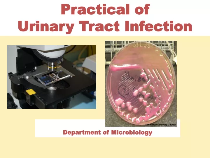
Practical of Urinary Tract Infection. Department of Microbiology. Important aspects of Microbiologic Examination of UTI: Urine collection - Urine analysis - Interpretation of microbiology laboratory result. Type of Specimens. Midstream urine (MSU) Clean catch Adhesive bag

E N D
Practical of Urinary Tract Infection Department of Microbiology
Important aspects of Microbiologic Examination of UTI: • Urine collection - Urine analysis - Interpretation of microbiology laboratory result
Type of Specimens • Midstream urine (MSU) • Clean catch • Adhesive bag • Suprapubic Aspiration • Catheter sample
The urinary catheter Urine specimens for laboratory investigations can be collected from catheterized patients as shown (left). The second port is for putting fluids into the bladder (right). Urine from the drainage bag should not be tested because it may have been standing for several hours.
TRANSPORT MEDIA Sterile Urine container
Urine analysis; 1- Dip stick (leukocyte esterase ,nitrate test) 2-microscopic ex; cell-counting chamber
Urine analysis; 1- Dip stick (leukocyte esterase ,nitrate test)
culture media blood agar MacConkey agar CLED agar an enriched medium Selective medium a differential medium
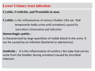
Laboratory examination of urine Quantitative (Colony counts) a urine sample is streaked on surface of Blood Agar plate and CLED agar / Mc Conkey agarwith a special loop calibrated to deliver a known volume. + 1 + 2 + 3 + 4 Over night incubation Isolation of colonies, Biochemical tests, Drug susceptibility test, Over night incubation RESULT Urinary Tract infection Module’05 …..
Other organisms ; • Candida albicans • Schistosoma haematobium • Tricomonas vaginalis
MacConkey's agar showing both lactose and non-lactose fermenting colonies. Lactose fermenting colonies are pink whereas non-lactose fermenting ones are colourless or appear same as the medium.|
CLED agar Selective culture medium for detection and isolation Of Escherichia coliandcoliformbacteria in urine
MacConkey’s agar plate showing growth of:Lactose fermenter pink coloniese.g. E. coli
E coli Indole Reactions Negative Positive
MacConkey’s agar plate showing growth of:Lactose fermenter pink coloniesKlebsiella
MacConkey’splate showing growth of: Non - Lactose fermenter pale coloniese.g. Proteus
Urease Test proteus is Urease positive Urease splits urea into ammonia; and alkalinizes the urine with production of crystals
Proteus growth : Swarming CLED (Cystine-Lactose-Electrolyte-Deficient) - inhibits the proteus swarm
Nutrient agar plate showing growth of:Blue-greencoloniesPseudomonas
Three API 20E strips : • Immediately after inoculation • After 24 hours incubation • That in ( b) after addition of reagents to certain wells. The organisms here is Escherichia coli. Here the first carbohydrate well (glucose) is also used for the nitrate reduction test
Biochemical Identification Enterococcus species • Bile Esculin hydrolysis Both Group D streptococci and enterococci produce a positive (left) bile Esculin hydrolysis test.
MICROSCOPIC APEARANCEGram positive cocci in clusters most likely staphylococci FROM CULTURE SMEAR FROM SPESIMEN: Pus cells & Gram positive cocci in clusters
To differentiate between Staphylococcus aureus & Staphylococcus epidermidis use the following test: • 1.COAGULASE TEST: • Tube coagulase test • Slide coagulase test • 2. DNAaseTEST • 3.MANITOL FERMINTATION TEST
Blood agar plate showing growth of :Staphylococcus aureusColonies are golden yellow in color
Staphylococcus spp Staphylococcus aureus Staphylococcus epidermidis Golden colonies (yellowish) white colonies
CATALASE TEST Procedure: Mix the colony in a drop of hydrogen peroxide (H2O2)
Differential Characteristics Catalase 2H2O2 O2 + 2H2O Streptococcivs. Staphylococci
Staphylococcus aureus S. aureus Coagulase POS Coagulase NEG Differential Characteristics
NOVOBIOCIN TEST Staphylococcus saprophyticus (resistant-Novobiocin) Staphylococcus epidermidis (sensitive-Novobiocin )
Gentamisin (CN) : 12 - 15 Chloramphenicol (C) : 12 - 18 Penicilin (P) : 28 - 29 R (Resistant) ; S (Sensitive) P P C C Staphylococcus aureus CN CN
Case 1 • The blood agar plate and CLED plate provided were inoculated with a sample of urine from a patient with a suspected urinary tract infection. Examine the plates and photographs provided. • Identify the colonies on the blood agar plates and photographs. • The photographs show the results of the Gram stain of each colony type. • Large colonies are Gram……….and small colonies are Gram………. CLED plate Blood agar Gram stain
Case 2 These Blood agar and CLED agar plates were inoculated with MSU from a 45 years old man suspected of having bladder stone and complaining of burning micturation. Urine examination showed : Moderate number of WBC and a PH of 8 CLED Blood agar A) What is the likely this pathogen? B) How would you confirm the identity of this pathogen? C) What is the role of this organism in forming stones?
Candida albicans Growth on Sabouraud's Dextrose Media Candida albicans on blood agar;