Download
1 / 68
680 likes | 1.16k Views
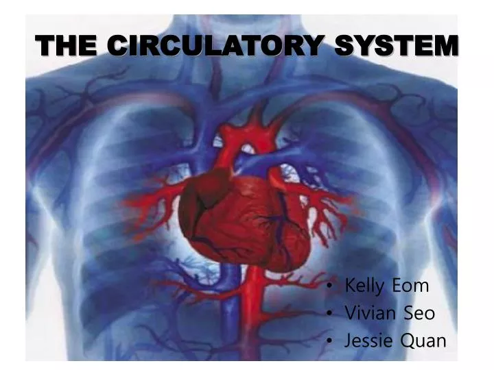
THE CIRCULATORY SYSTEM. Kelly Eom Vivian Seo Jessie Quan. Circulatory system. Diffusion is inefficient over long distances. Circulatory system rapidly transport fluid throughout the body. Main functions - exchange gases - absorb nutrients - dispose of wastes. Circulatory system.

E N D
THE CIRCULATORY SYSTEM • Kelly Eom • Vivian Seo • Jessie Quan
Circulatory system Diffusion is inefficient over long distances. Circulatory system rapidly transport fluid throughout the body. Main functions - exchange gases - absorb nutrients - dispose of wastes
Circulatory system Open circulatory system: -It pumps blood into an internal cavity called sinuses, which bathe tissues with an oxygen- and nutrient-carrying fluid called hemolymph. -The hemolymph returns to the pumping mechanism of the system, a heart, through holes called ostia. -It occurs in insects and most mollusks.
Circulatory system Closed circulatory system: -The nutrient-, oxygen-, and waste-carrying fluid, blood, is confined to vessels. -It is found among earthworms, mollusks, and vertebrates. - Humans and other vetebrates have a closed circulatory system specifically called, cardiovascular system.
Cardiovascular system Please put this slide in slide show. Arteries- vessels moving away from the heart/ oxygenated blood Arterioles- branched smaller vessels of arteries Capillaries- smallest vessels/ gas and nutrient exchange Capillary beds- networks of capillary vessels Venules-vessels returning to the heart/ deoxygenated blood Veins- larger vessels of venules
Blood flow 1. Oxygen-poor blood from the body flows into the right atrium (on the right side of heart) through two veins, the upper superior(anterior) vena cava and the lower inferior(posterior) vena cava. 2. The blood moves through the right atrioventricular valve(AV valve) and flows into the right ventricle. 3. Right ventricle pumps blood to the lungs via the pulmonary arteries. 4. As blood flows through capillary beds in the left and right lungs, it releases waste gases and picks up oxygen. 5. This newly oxygen-rich blood returns from the lungs to the left atrium through the pulmonary veins. 6. The blood flows through the left atrium into the left ventricle as ventricle opens and the atrium contracts.
Blood Flow 7. The left ventricle pumps the oxygen-rich blood out through the aorta, which conveys blood to arteries leading throughout the body. 8. Branches from aorta lead to capillary beds in the head and arms. 9. The aorta continues to posterior direction, in abdominal organs and legs. 10. Oxygen-poor blood from the head, neck, and forelimbs is channeled into superior vena cava. 11. Blood from the trunk and hind limbs is drained by the inferior vena cava. 12. The blood goes into the right atrium.
Heart About the size of a clenched fist Consists mostly of cardiac muscle Two atria: have thin walls and serve as collection chamber for blood returning to the heart Ventricles: have thicker walls and contract more strongly than the atria - left ventricle: must pump blood to all body organs→ strong contraction
Heart The systemetic circuit: the circulation pathway throughout the body The pulmonary circuit: the blood pathway between the right side of the heart, to the lungs, and back to the left side of the heart
The cardiac cycle The cardiac cycle: one complete sequence of pumping and filling - Systole: the contraction phase - Diastole: the relaxation phase The cardiac cycle is regulated by → autorhythmic cells : specialized tissues in the heart : self-excitable and able to initiate contractions without external stimulation by nerve cells. Cardiac output- the volume of blood per minute that the left ventricle pumps into the systemic circuit - depends on : 1. heart rate- the rate of contraction 2. stroke volume- the amount of blood pumped by the left ventricle in each contraction.
The cardiac cycle Four valves in heart -Consist of flaps made of connective tissue -Prevent backflow -Keep blood moving in the correct dirrection Atrioventricular(AV) valves: between each atrium and ventricle - Anchored by strong fibers that prevent them from turning inside out - Pressure generated by the ventricles closes AV valves Semilunar valves: where the aorta leaves the left ventricle/ the pulmonary artery leaves the right ventricle - Forced open by pressure generated by contraction of ventricles Pulse: the rhythmic stretching of arteries caused by the pressure of blood driven by the powerful contractions of the ventricles Heart murmur - result of defect in one or more valves - Detectable as a hissing sound when a stream of blood squirts backward through a valve
Heart beat The cardiac cycle occurs as The SA(sinoatrial) node, or pacemaker, located in the upper wall of the right atrium, initiates the cycle -by simultaneously contracting both atria -by sending a delayed impulse that stimulates the AV(astrioventricular) node.
Heart beat The AV node in the lower wall of the right atrium sends an impulse through the bundle of His, nodal tissue that passes down between both ventricles and then branches into the ventricles through the Purkinje fibers. - This impulse results in the contraction of the ventricles.
Heart beat 3. →When the ventricles contract(the systole phase) - blood is forced through the pulmonary arteries and aorta. - the AV valves are forced to close as well. → When the ventricles relax(the diastole phase) - backflow into the ventricles causes the semilunar valves to close. - The closing of AV valves, followed by the closing of the semilunar valves, produces the characteristic “lub-dup” sounds of the heart.
Blood • Volume of flow per second must be constant through the entire pipe. • Blood is constantly flowing through out bodies.
Volume of flow per second must be constant through the entire pipe. • Since volume of flow per second must be constant through entire pipe, fluid must flow faster as cross-sectional are of pipe narrows. • The total cross-sectional are of capillaries determines flow rate. • Blood travels over a thousand times faster in aorta. • Velocity change follows from the law of continuity, which describes fluid movement through pipes.
A fluid flows through narrower segments of the pipe faster than it flows through wider segments.
Blood is constantly flowing through out bodies • Blood is pumped by the heart. • Blood travels through blood vessels. • Blood Carries nutrients, water, oxygen, and waste products. • Red blood cells, white blood cells, platelets, and plasma are in the blood.
Blood is pumped by the heart. • Heartbeat gives pressure to blood. • Blood pressure is the main force driving blood from the heart to the capillaries. • Blood pressure, the hydrostatic force exerted against wall of blood vessel. • Blood pressure is highest in the arteries during systole which is called systolic pressure. • Blood flow velocity decreases markedly in arterioles and is lowest in capillaries. • Hydrostatic pressure drives blood from heart to capillary beds.
Peripheral resistance which is caused by narrow openings of arterioles impeding the exit of blood from arteries causes swelling of arteries during systole. • Recoiling of stretched elastic arteries during diastole creates diastolic pressure. • Diastolic pressure maintains continuous blood flow into arterioles and capillaries.
Blood travels through blood vessels. • Blood leaves the heart from left ventricle to the aorta which is the largest artery in the body. • Blood that leaves the aorta has a lot of oxygen which is important for cells to do their function. • Oxygen rich blood travels throughout arteriesinto smallest arterioles. • Blood travels through capillaries to vein to go back to the heart.
When blood reaches lungs, carbon dioxide is removed from blood and replaced with fresh oxygen that had inhaled through lungs. • Blood flows through vessels of circulatory system at uneven speeds and pressures.
Arteries • Arteries have three layers. • On outside, a layer of connective tissue with elastic fibers allows vessel to stretch and recoil. • Middle-layer contains smooth muscle and more elastic fibers.
Lining the lumen of all blood vessels, an endothelium, is a single layer of flattened cells that provides a smooth surface that minimizes resistance to flow of blood. • Blood vessels that carry oxygen rich blood. • Arteries transport blood away from heart.
Arteries carry bright red blood which color comes from oxygen that it carries. • Arteries have thicker middle and outer layers than veins. • Thicker walls of arteries provide strength to accommodate blood pumped rapidly and at high pressure by heart. • Thinnest arteries are called arterioles.
Capillaries • Capillaries are tiny and thin blood vessels. • capillaries connect arteries to veins. • Nutrients, oxygen, and wastes pass in and out of blood through capillary walls.
Capillaries’ thin walls consist only of endothelium and its basement membrane. • Total cross-sectional area is much greater in capillary beds than in any other part of circulatory system. • Capillaries are only vessels with walls thin enough to permit the transfer of substances between the blood and interstitial fluid. • The slower flow of blood through tiny vessels enhances exchange.
When blood leaves the capillaries and enters venules and veins, it speeds up reduction in total cross-sectional area. • During strenuous exercise, blood is diverted from digestive tract and supplied more generously to skeletal muscles and skin. • Two mechanisms regulate distribution of blood in capillary beds. • Both depend on smooth muscles controlled by nerve signals and hormones.
In one mechanism, contraction of smooth muscle layer in wall of an arteriole constricts vessel, reducing its diameter and decreasing blood flow through it to a capillary bed. • In other mechanism, rings of smooth muscle called precapillary sphincters which are located at entrance to capillary beds control flow of blood between arterioles and venules. • Exchange of substances between blood and interstitial fluid that bathes cells takes place across thin endothelial walls of the capillaries. • Small molecules, like oxygen and carbon dioxide, diffuse down concentration gradients across endothelial cells.
Diffusion can occur through clefts between adjoining cells. • Blood pressure within capillary pushes fluid through capillary clefts. • Blood cells suspended in blood and most proteins dissolved in blood are too large to pass readily through endothelium and remain in capillaries. • Blood proteins remaining in capillaries create constant osmotic pressure from arteriole to venule end of a capillary bed. • Difference between blood pressure and osmotic pressure drives fluids out of capillaries at the arteriole end and into capillaries at the venule end.
Lost fluid and proteins return to blood via the lymphatic system. • The fluid called lymph is in lymphatic system; its composition is about same as interstitial fluid. • Lymph vessels have valves that prevent backflow of fluid toward capillaries. • Along a lymph vessel are organs called lymph nodes. • When body is fighting an infection, lymph nodes become swollen and tender. • Lymphatic system helps defend against infection and maintains volume and protein concentration of blood.
Vein • Arteries have three layers. • On outside, a layer of connective tissue with elastic fibers allows vessel to stretch and recoil. • Middle-layer contains smooth muscle and more elastic fibers.
Lining the lumen of all blood vessels, an endothelium, is a single layer of flattened cells that provides a smooth surface that minimizes resistance to flow of blood. • Veins carry oxygen poor blood back to heart. • Smallest veins, venules, join larger veins that open into heart. • Thinner-walled veins convey blood back to the heart at low velocity and pressure. • Blood in the vein flows mainly by muscle actions.
Skeletal muscles squeeze veins and push blood through them. • Flaps of tissue within veins act as one-way valves that allow blood to flow only toward the heart.
Blood Carries nutrients, water, oxygen, and waste products. • Nutrients, water, oxygen, and waste products are carried by blood cells in blood vessel to transport to and from body cells.
Red blood cells, white blood cells, platelets, and plasma are in blood • Red blood cells • White blood cells • Platelets • Plasma
Red blood cell • Red blood cells carries oxygen and carbon dioxide. • Red blood cells pick up oxygen in lungs and transport it to all body cells. • Red blood cells gather up carbon dioxide after delivering oxygen to cells.
Red blood cells transport carbon dioxide back to lungs where it is removed from body when human exhales. • About 5,000,000 red blood cells are in one drop of blood. • Red blood cells are heavier than other blood cells. • Red blood cells are made in bone marrow which is a soft tissue inside of bone. • Red blood cells are also called erythrocytes.
The small size and biconcave shape of erythrocytes provide a large collective surface area for the total population of erythrocytes. • The greater of total area of erythrocyte membrane in a volume of blood leads to more rapid oxygen diffusion. • Hemoglobin are iron-containing protein that transports oxygen. • Erythrocytes lack nuclei and mitochondria and generate AP exclusively by anaerobic metabolism.
White blood cells • White blood cells are also called leukocytes. • White blood cells help body fight off germs. • White blood cells attack and destroy germs when they enter the body.
Body produces more white blood cells if infection exists in the body. • White blood cells produce in bone marrow. • Blood contains five major types of white blood cells which are monocytes, neutrophils, basophils, eosinophils, and lymphocytes. • Monocytes and neutrophils are phagocytes which engulf and digest bacteria and debris from body’s own dead cells. • Lymphocytes develop into specialized B cells and T cells which produce immune response against foreign substances.
White blood cells spend most of time outside the circulatory system, patrolling through interstitial fluid and lymphatic system, where most of battles against pathogens are waged.
Platelets • Platelets are blood cells that help stop bleeding. • As platelets stick to opening of damaged vessel they attract more platelets, fibers, and other blood cells to help form a plug to seal broken blood vessel.
The wound stops bleeding when platelet plug is completely formed; the platelet pugs are called scabs. • Platelets produce in bone marrow. • Platelets have no nuclei and originate as pinched-off cytoplasmic fragments of large cells in bone marrow.
Stem cells • Erythrocytes, leukocytes, and platelets all develop from a common source. • A single population of cells called pluripotent stem cells in red marrow of bones, particularly ribs, vertebrae, breastbone, and pelvis.
A negative- feedback mechanism, sensitive to amount of oxygen reaching body’s tissues via blood, controls erythrocyte production. • If tissues do not receive enough oxygen, kidney synthesizes and secretes a hormone called erythropoietin which stimulates production of erythrocytes. • If blood transport excessive oxygen, level of EPO falls and erythrocyte production slows.
Plasma • Plasma is the liquid part of the blood. • Approximately half of blood is made of plasma. • The plasma carries blood cells and other components throughout body.
Plasma is made in liver. • Plasma is lighter than other blood cells. • Plasma is 95% of water and other 5% of dissolved substances including salts.