Download
1 / 22
360 likes | 858 Views
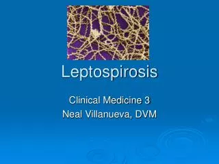
Leptospirosis. Sung Chul Hwang Dept. of Pulmonary and Critical Care Medicine Ajou University School of Medicine. Introduction. Spirochetal disease, finely coiled, motile, 0.1 m x 6 – 20 m Systemic infection manifested as widespread vasculitis Zoonosis

E N D
Leptospirosis Sung Chul Hwang Dept. of Pulmonary and Critical Care Medicine Ajou University School of Medicine
Introduction • Spirochetal disease, finely coiled, motile, 0.1 m x 6 – 20 m • Systemic infection manifested as widespread vasculitis • Zoonosis • L. interogans 23 serogroups and 187 serovars • L. biflexa : non-pathogenic, saprophyte
Historical back ground • 1921 : Takaki 창경원 죽은 족제비 – L. icterohemorragiae 분리 • 1942 : sekiguchi – L. canicola from mouse • 1951 : 미군, 동경 401 의무 시험소-국내 들쥐로부터 ictohemorrhagiae 분리 • 1975 : 경기 강원 충북, “출혈성 폐렴양 괴질” • 1984년 10월: 강원도 원주, 괴질 환자에서 렙토스피라 균을 분리 동정 • 1984년이후: 매년 9-11월 환자 및 야생쥐-leptospira 균이 동정됨
Epidemiology • Disease of the wild animals • Incidental human infection by direct or indirect contact with the animal • 20-40s active males: farmers or soldiers in harvest time • 9-10 peak into November • 추수, 탈곡, 벌초, 성묘, 나무하기, 훈련, 등
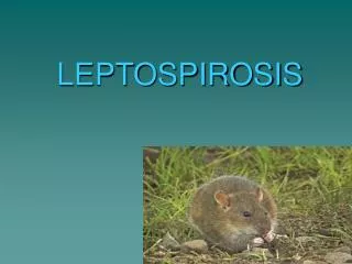
Reservoires of Infection • Rats • Dogs • Live stocks • Rodents including rabbits • Wild animals • Cats
Sources of Human Infections • Contaminated Water or soil from infected urine • Direct animal contacts • Occupational exposure : farmers, vets, abattoire workers • Recreational exposure : campers, swimmers, visiting graveyards
Routes of Infection • Contact with water or soil contaminated animals • Direct contact with the by urine from infected source, farmer, vets, butchers, recreational activities • Rodents carry EH fever, scrub typhus, paratyphus, leptospirosis • Factors for high incidence : rain during harvest time, carrier rate in rodents • Spirochetes survive longer in wet swampy conditions
국내 주민의 항체 보유율 • 1985.2 – 1986.7 : 11.69% • 1987.2 – 1987. 7 : 5.9 % • 1985 in febrile patients : 20% • 1986- 1987 in febrile patients : 11.6%
국내 야생쥐의 균 보유율 • 1984 : 15.5% • 1985 : 14.9% • 1986 : 16% • 1987 : 30.9% ( 파주, 여주)
Microbiology and distribution • Mainly serogroup ictohemorrhagiae and canicola • 전북, 서울, 강원, 충북, 충남 • CH-48 : 춘천지방, 혈청형 미상 • Serovar : mainly lai
Pathogenesis • Entry sites : skin wounds or abrasions in hand and feet and mucous membranes, conjunctiva, nasal, oral • Bacteremia involving the entire body including eye, CSF • Systemic effect and vasculitis due to endotoxin (hyaluronidase) and burrowing motility • Hemorrhagic necrosis esp. in liver, lung, and kidneys jaundice, ARF, hemorrhages
Phase I (Septicemic) • Following incubation period of 7-10 days • High spiking fever, headaches, myalgia, arthralgias • Lasting 4 – 7 days • Proteinuria and increased creatinnine • Organism detectable but serologic diagnosis not possible
Phase II (Immune) • Much more variable • Induction of IgM Antibodies • 1- 3 day freedom recurrence of symptoms • Lower fever, CNS signs • Maybe cultured from urine but not from blood or CSF
Weil’s Disease • Less common but severe form • Mild phase I, initially • Followed by severe Jaundice , Azotemia, and Hemorrhage from Lungs, GI tract, and other organs (3-6 day) • Oliguric renal failure and Liver dysfunction dominate the clinical picture
Clinical Signs of Leptospirosis • Pulmonary infiltrates, pneumonitis, hemorrhages • Conjunctival injection • Jaundice • Muscle tenderness • Abdominal tenderness • CVA tenderness • Abnormal auscultation • Erythema, petechiae, neck stiffness, adenopathy
Lab. Diagnosis • Microbiologic identification : Blood or CSF first 10 days Urine second week (Fletcher’s, EMJH Medium) • Serology: screeningMicroscopic Slide Agglutination (MST), titration & serogroup identification Microscopic Agglutination (MAT), detection of IgM (ELISA)
Chest X-rays • 33 – 64 % of patientssjows abnormality • Bilateral nodules, rosette densities • Diffuse ill-defined infiltrates • Massive confluent consolidation • Bilateral, Non-lobar, peripheral predominance • Rare pleural reaction • Complete resolution within 5 to 10 days
Treatment • Early anti-microbial therapy is importantshorten the course and prevent carrier state • Choice : Penicillin G, Ampicillin • May cause “ Jarish-Huxheimer type reaction” • Mild cases oral Doxycycline or Amoxicillin
Prevention • Vaccination of domestic animals • Rodent control • Protective gloves and boots • Avoid swimming in contaminated waters • Vaccination in endemic region
Differential Diagnosis • EH fever • Rickettsial disease : Scrub typhus, murine typhus • Acute viral hepatitis • Sepsis • Influenza • Aseptic Meningitis