Download
1 / 37
370 likes | 509 Views
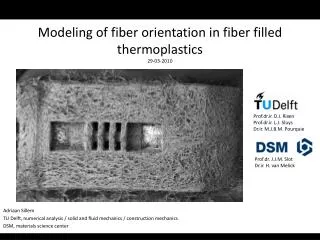
Modeling cancer (and mechanics). Lecture 7 of Introduction to Biological Modeling Nov. 10, 2010. Steve Andrews Brent lab, Basic Sciences Division, FHCRC. Modeling mechanics Cancer introduction Cancer incidence models Tumor growth models Summary. Alberts and Odell: Lysteria motility.

E N D
Modeling cancer (and mechanics) Lecture 7 of Introduction to Biological Modeling Nov. 10, 2010 Steve Andrews Brent lab, Basic Sciences Division, FHCRC
Modeling mechanics Cancer introduction Cancer incidence models Tumor growth models Summary
Alberts and Odell: Lysteria motility Credits: Alberts and Odell, PLoS Biology, 2:e412, 2004.
Nédélec: microtubule asters Q: What stabilizes the microtubule asters? A: double-headed motors that push and pull. movies: http://www.embl.de/~nedelec/reprints/asters/figure2/index.html Credits: http://dms.dartmouth.edu/compton/photos/photos/#; Nédélec, J. Cell Biol. 158:1005, 2002.
Odell and Foe: eukaryotic cell division furrow After DNA segregation, dynamic microtubules extend in all directions, and stable ones extend towards equator. MKLP1 motors carry centralspindlin to cortex at equator, which activates Rho, which activates actin and myosin, which contracts the cell. Credit: Odell and Foe, J. Cell Biol. 183:471, 2008.
Wolgemuth: myxobacteria gliding with slime load-velocity curve slime nozzle Slime expands when it leaves the cell because it hydrates. Expansion creates a force that pushes the bacterium forwards. Credits: Wolgemuth et al. Current Biology 12:369, 2002.
Andrews and Arkin: shapes of bacterial polymers Many bacteria have helical or ring-shaped membrane-bound polymers. These shapes can arise from simple mechanics. different parameters create different shapes Credit: Andrews and Arkin, Biophys. J. 93:1872, 2007.
Overview of cellular mechanics modeling Lots of research on polymers • actin, microtubules (and motors) • bacterial cytoskeletal polymers • DNA, RNA, nuclear pore polymers, etc. Other mechanics research • cell motility with slime • membrane shape • development, growth, gastrulation, wound healing Methods • physics: mechanics, polymer & membrane physics, rheology • custom software (MatLab, C, C++, Java, etc.) • many “agent-based” models Good book Jonathon Howard, Mechanics of Motor Proteins and the Cytoskeleton, 2001.
Modeling mechanics Cancer introduction Cancer incidence models Tumor growth models Summary
Cancer Cancer is important • kills 1/5 of Americans • somewhat preventable (e.g. smoking, obesity, UV radiation, screening) • somewhat curable (e.g. surgery, chemotherapy, radiation) • 4.8 billion $/year NCI funding • mission of the Hutch, and other cancer centers Cancer is complex • can arise in any organ or tissue • causes include: mutations, epigenetic mutations, viruses • oncogenes and tumor-suppresor genes • cell systems: signaling, cell cycle, DNA repair, apoptosis • stages: DNA damage, proliferation, vascularization, metastatis
Cancer modeling Lots of statistical modeling • identifying cancer causes • finding tumor suppresor genes and oncogenes • analysis of cancer incidence rates Some tumor development modeling Surprisingly little biochemical modeling Some cancer modeling resources Center for the Development of a Virtual Tumor https://www.cvit.org Cancer Intervention and Surveillance Network http://cisnet.cancer.gov
Colorectal cancer Typical steps mutation that inactivates APC/-catenin pathway mutation in CDC4 and other cell cycle genes, causing chromosomal instability mutations in KRAS/BRAF oncogenes (EGF signaling pathway) additional mutations invasion of tumor into underlying tissues (a carcinoma) metastasis to other parts of the body Credit: Jones et al.Proc. Natl. Acad. Sci. USA 105:4283, 2008.
Colorectal cancer Credit: Jones et al.Proc. Natl. Acad. Sci. USA 105:4283, 2008.
Modeling mechanics Cancer introduction Cancer incidence models Tumor growth models Summary
Cancer incidence questions Why study cancer incidence? • improve understanding of cancer How long does each step take? How many mutations are required? • find best time(s) for cancer screening tests • predict outcomes for individuals • risk assessment • predict benefit of an intervention
Raw data, from 1950s Note log-log axes All lines are y=ct6 Credit: Armitage and Doll, Int. J. Epidemiology, 33:1174, 2004, which is reprint of Armitage and Doll, Br. J. Cancer 8:1, 1954.
Armitage and Doll’s theory incidence ~ t6 could arise from • 7 rare events • in a specific sequence • each event has a constant liklihood over time
Armitage and Doll math Suppose probability of occurance of rth event is pr per unit time So, probability that 1st event has happened is about p1t probability that 1st event has happened time (age) Ignoring the sequence of events, the probability of event #1, and event #2, and ..., and event #6 is (p1t)(p2t)(p3t)(p4t)(p5t)(p6t) = p1p2p3p4p5p6t6 There are 6! possible sequences of 6 events. Only one of them is the “correct” one. So, the probability that 6 events have happened, in the correct sequence, is
cancer death rate = Armitage and Doll math The probability of the 7th event occuring during time interval ∆t is p7∆t. The probability that 6 events happened by time t, and then the 7th event during the next ∆t is For r events: Why a specific sequence? (death rate is still ~t6 if the sequence is ignored) • data show that cancer probability is directly proportional to carcinogen concentration • data show a long lag time between carcinogen exposure and cancer • These makes sense if the carcinogen only affects event #1.
Improved cancer incidence model Problems with Armitage-Doll model • the exact A-D model suggests ~10 sequential rare events, not 7 • biology work suggests only about 2 or 3 necessary rare events (APC/-catenin mutations) • newer and better data don’t fit t6 incidence curve Incidence of colorectal cancer, 1975 - 2004 Note linear scales, not log-log scales Credit: Meza et al. Proc. Natl. Acad. Sci. USA 105:16284, 2008.
Multi-stage clonal expansion (MSCE) model 3 stage model (Luebeck’s group) • Initiation requires 2 rare events mutation of both copies of APC tumor suppresor, rates are 0 and 1 • Clonal expansion of initiated cells cell division rate , cell death or differentiation rate net cell growth rate – • One of the new cells transforms to metastatic malignant transformation rate 2 Credit: Meza et al. Proc. Natl. Acad. Sci. USA 105:16284, 2008.
MSCE model results Colorectal cancer Pancreatic cancer Credit: Meza et al. Proc. Natl. Acad. Sci. USA 105:16284, 2008.
MSCE model interpretation X slope = X01 exponential growth rate = – adenoma sojourn time 0 Credit: Meza et al. Proc. Natl. Acad. Sci. USA 105:16284, 2008.
MSCE model application When is the best age for a colonoscopy screening? Answer: if 1 screen: age 57 if 2 screens: ages 50 and 68 Credit: Jeon et al. Mathematical Biosciences 213:56, 2008.
MSCE model conclusions Model fits incidence data Model agrees with biology • 2 rate-limiting mutations • then, chromosomal instability, so more mutations follow • time for growth of adenoma • rate-limiting transformation to metastatic • fast cancer after metastatic Model enables screening recomendations
Modeling mechanics Cancer introduction Cancer incidence models Tumor growth models Summary
No angiogenesis Glazier’s tumor growth model Day 30: cylindrical tumor Day 15: spherical tumor with 200 m diameter pre-existing vasculature Day 75: tumor has ruptured blood vessels green = normal tumor cell yellow = hypoxic tumor cell blue = necrotic tumor cell Credits: Shirinifard et al. PLoS ONE 4:e7190, 2009.
Movie, no angiogenesis Credits: Shirinifard et al. PLoS ONE 4:e7190, 2009.
Glazier’s results, with angiogenesis pre-existing vasculature Day 15: spherical tumor with 300 m diameter Day 30: cylindrical tumor Day 75: developed vascularized tumor green = normal tumor cell yellow = hypoxic tumor cell blue = necrotic tumor cell purple = new vasculature Credits: Shirinifard et al. PLoS ONE 4:e7190, 2009.
Glazier’s results, with angiogenesis Credits: Shirinifard et al. PLoS ONE 4:e7190, 2009.
Quantitative results all tumor cells normal (not hypoxic) tumor cells Credits: Shirinifard et al. PLoS ONE 4:e7190, 2009.
GGH / cellular Pott’s model Each lattice site is • empty (i.e. extracellular medium) • normal tumor cell (green) • hypoxic tumor cell (yellow) • necrotic cell (blue) • vascular cell (red) • neovascular cell (purple) The system “energy” depends on • contact energy at each cell-cell contact face • pressure energy for compressed cells In each “move”, the simulator • randomly changes the contents of a random site • accepts the move if it lowers the system energy • accepts the move with a low probability if it raises the system energy • otherwise, rejects the move and returns to the prior state
GGH / cellular Pott’s model More rules • cells are normal or hypoxic, depending on oxygen availability • cells grow (target volume increases), depending on oxygen availability • cells divide if their volume exceeds the “doubling volume” • necrotic cells shrink (target volume decreases) • hypoxic cells secrete VEGF-A (vascular endothelial growth factor) • vascular cells secrete chemoattractant • neovascular cells grow towards chemoattractant using another “energy” function
GGH / cellular Pott’s model Each lattice site has • oxygen concentration • VEGF-A concentration (vascular endothelial growth factor) • chemoattractant for vascular growth These are modeled with reaction-diffusion equations VEGF equation: decay rate of VEGF-A production of VEGF-A by hypoxic cells diffusion of VEGF-A equations are similar for oxygen and chemoattractant
Software CompuCell3D http://www.compucell3d.org 3D reaction-diffusion simulations cellular Potts model simulations used for simulating • morphogenesis • tumor growth • cell sorting • biofilms • foams (?)
Summary Mechanics • polymer models • slime extrusion model Cancer incidence • Armitage-Doll model (incidence ~ t6) • Luebeck model (incidence has lag, then linear) Tumor growth • with and without angiogenesis • cellular Potts model • reaction-diffusion model • CompuCell3D software
Course summary Introduction Modeling dynamics Metabolism Gene regulatory networks Stochasticity and robustness Spatial modeling Mechanics and cancer